

ABSTRACT
Using engineered initiator tRNA for precise control of protein translation within cells has great promise within future orthogonal translation systems to decouple housekeeping protein metabolism from that of engineered genetic systems. Previously, E. coli strain C321.ΔA.exp lacking all UAG stop codons was created, freeing this ‘amber’ stop codon for other purposes. An engineered ‘amber initiator’  that activates translation at UAG codons is available, but little is known about this tRNA’s orthogonality. Here, we combine for the first time the amber initiator
  in C321.ΔA.exp and measure its cellular effects. We found that the
  expression resulted in a nearly 200Yfold increase in fluorescent reporter expression with a unimodal population distribution and no apparent cellular fitness defects. Proteomic analysis revealed upregulated ribosomeYassociated, tRNA degradation, and amino acid biosynthetic proteins, with no evidence for offYtarget translation initiation. In contrast to previous work, we show that UAGYinitiated proteins carry NYterminal methionine exclusively. Together, our results identify beneficial features of using the amber initiator
  to control gene expression while also revealing fundamental challenges to using engineered initiator tRNAs as the basis for orthogonal translation initiation systems.
The ability to precisely control an orthogonal protein translation system within engineered organisms is one of the grand goals of synthetic biology.1 A distinct feature of the standard genetic code is the dual function of AUG as both the dominant start codon as well as a methionine sense codon internal to genes. This dual function of the AUG start codon can create ambiguities in gene prediction and genetic design. For example, in recombinant protein design, inYframe AUG sense codons can behave as start codons if the RNA sequence upstream resembles a ShineYDalgarno site.2 Therefore, engineering an orthogonal system to initiate translation at a unique codon reserved solely for that function would be beneficial. Once established, the unique start codon could be paired with an orthogonal initiator tRNA aminoacylated with natural or unnatural amino acids with useful properties as has been done before in internal codons.3
Specific tRNAs recognise and decipher mRNA codons during translation and sequentially associate a bound amino acid to a growing polypeptide within the ribosome through three anticodon nucleotides (positions 34Y36) on the anticodon loop. tRNAs are divided into two functionally and structurally distinct classes: initiator tRNAs and elongator tRNAs. Initiator tRNAs participate in the initiation phase of translation, recognizing start codons, and thus, incorporate the first amino acid. The three consecutive G:C base pairs in the tRNA anticodon stem is a crucial conserved element for initiator tRNA binding to the PYsite in the 30S ribosomal subunit via interactions with the 16S ribosomal RNA.4Y5 Additionally, the initiator tRNA’s conserved C1:A72 base pair mismatch in the acceptor stem specifically interacts with methionylYtRNA NY formyltransferase enzyme6, and initiation factor 2 (IF2)7, a protein vital for prokaryotic translation initiation.
Over the past two decades, efforts have focused on engineering stop codon suppression systems that use elongator tRNA aminoacylYsynthetase pairs as an orthogonal system that specifically recognises amber (UAG) sense codons to siteY specifically incorporate unnatural amino acids 8. Unnatural amino acid incorporation enables researchers to build engineered proteins with an extended range of structural and functional properties for applications in protein evolution9, therapeutics10, and elucidating structure and function11. To prevent undesired natural amber codon suppression, competition for UAG codons from release factor 1, and nonYspecific unnatural amino acid incorporation, researchers have recently built a genomically recoded Escherichia coli strain, C321.ΔA with all instances of UAG stop codons and associated release factor (RF1) removed.12
The UAG amber stop codon has also been repurposed as a unique initiation codon in E. coli by creating an initiator  with anticodon changed from the canonical CAU to CUA. This amber initiator has been shown in chloramphenicol acetyltransferase (CAT) reporter assays on bulk cell cultures to initiate translation in vivo from a UAG start codon.13 They found that initiation efficiency was only 50Y60% relative to the AUG reporter and that it was aminoacylated predominantly by glutamine instead of methionine.13Y14 Despite excellent prior work characterizing the amber initiator tRNA, a more complete picture of its interactions with the host cell is needed if an orthogonal translation system is to be based on this tRNA.
In this work, we characterized the phenotypic effects of expressing the amber initiator  in the genomically recoded C321.ΔA.exp strain using a modular tRNA/reporter plasmid system. Using this system we found that expressing
  in E. coli C321.ΔA.exp resulted in initiation from a UAG start codon with similar populationYlevel characteristics to that seen from the canonical AUG initiation codon and wildYtype initiator tRNA. We also found previously uncharacterised effects on host proteome, but surprisingly, found no evidence for offYtarget translation initiation from genomic UAG codons. Additionally, we found that proteins initiated from
  carried methionine exclusively at their NYterminus. Together, this work reveals specific opportunities and challenges to using engineered initiator tRNAs in orthogonal translation systems.
RESULTS AND DISCUSSION
To measure orthogonality of the previously described amber initiator  we built a modular plasmidYbased amber initiator tRNA expression and reporter system (Figure 1A). The modular system consists of the amber initiator plasmid (pULTRA::tac2 metY(CUA)) (Figure 1A, top) harbouring the metY(CUA) gene expressing
  13 and a compatible reporter plasmid pQEY60::T5YsfGFP (Figure 1A, bottom) encoding one of three superfolder green fluorescent protein (sfGFP) variants differing in their start codon (either AUG, UAG, or GCC), where sfGFP(AUG) denotes the superfolder GFP gene with AUG start codon. To reduce host interference from release factor 1 competing with UAG codons we chose E. coli C321.ΔA.exp (Table 1) with a reduced genetic code as the host strain for this work.12
Description of strains and plasmids used in this study.

Expression of amber initiator tRNA causes increase in reporter fluorescence from both amber UAG and canonical AUG start codons. (A) Amber initiator and reporter plasmid system design. Amber initiator plasmid pULTRA::tac2metY(CUA) transcribing  , and reporter plasmid pQEY60 library expressing sfGFP containing three different initiation codons (AUG, UAG, and GCC). (B) Normalized expression levels from sfGFP reporters under different
  transcription conditions. Repressed, 2% glucose; Induced, 1 mM IPTG; tRNA, presence or absence of
  expressing amber initiator plasmid. Average of three biological replicates with error bars showing one standard deviation. (C) Representative fluorescence (Arb. U.) histogram of gated populations containing pULTRA::tac2metY(CUA) expressing
  with pQEY60 reporter library. Induced cell population (darker shades) and repressed cell population (lighter shades) shown for each reporter. Vertical dashed lines demarcate fluorescent and nonYfluorescent populations based on the 99th percentile of the control population (fluorescence of E. coli C321.∆A.exp population expressing
  with sfGFP(GCC) reporter).
We used this plasmid system to measure translation initiation events due to  expression. In the absence of pULTRA::tac2metY(CUA) plasmid, the sfGFP(UAG) reporter fluorescence was indistinguishable from the negative control sfGFP(GCC) fluorescence (Figure 1B). However, when the
  was present in the cell, sfGFP(UAG) reporter fluorescence increased 196Yfold in the induced condition compared to the repressed condition (Figure 1B and Figure S1). Moreover,
  expression resulted in sfGFP(UAG) fluorescence at a similar level to that of sfGFP(AUG) fluorescence when
  was absent in the system (Figure 1B and Table S1). Fluorescence from reporter sfGFP(GCC) remained nearly unchanged in all conditions, consistent with previous findings.15 These data confirm the ability of
  to efficiently initiate protein synthesis from the UAG stop codon at nearY cognate AUG levels.13
Surprisingly,  expression caused an increase in the sfGFP(AUG) fluorescence per cell (Figure 1B, right blue bars) suggesting the possibility that
  could also recognise and interact with AUG start codons. Previous work concluded that the wildYtype host initiator tRNA (with a CAU as opposed to CUA anticodon) does not initiate translation from a UAG start codon.15 The reciprocal interaction of a CUA anticodon with an AUG start codon should also result in weak translation initiation.
We next used flow cytometry to measure the distribution of fluorescence within the population of E. coli C321.ΔA.exp cells harbouring the pULTRA::tacYmetY(CUA) plasmid. All cell populations had unimodally distributed fluorescence in both repressed and induced conditions and had uniform populationYwide increase in fluorescence in both the sfGFP(UAG) reporter and the sfGFP(AUG) reporter strains upon  induction (Figure 1C). This is the first measurement, to our knowledge, of populationY level translationYinitiation from the amber initiator tRNA.
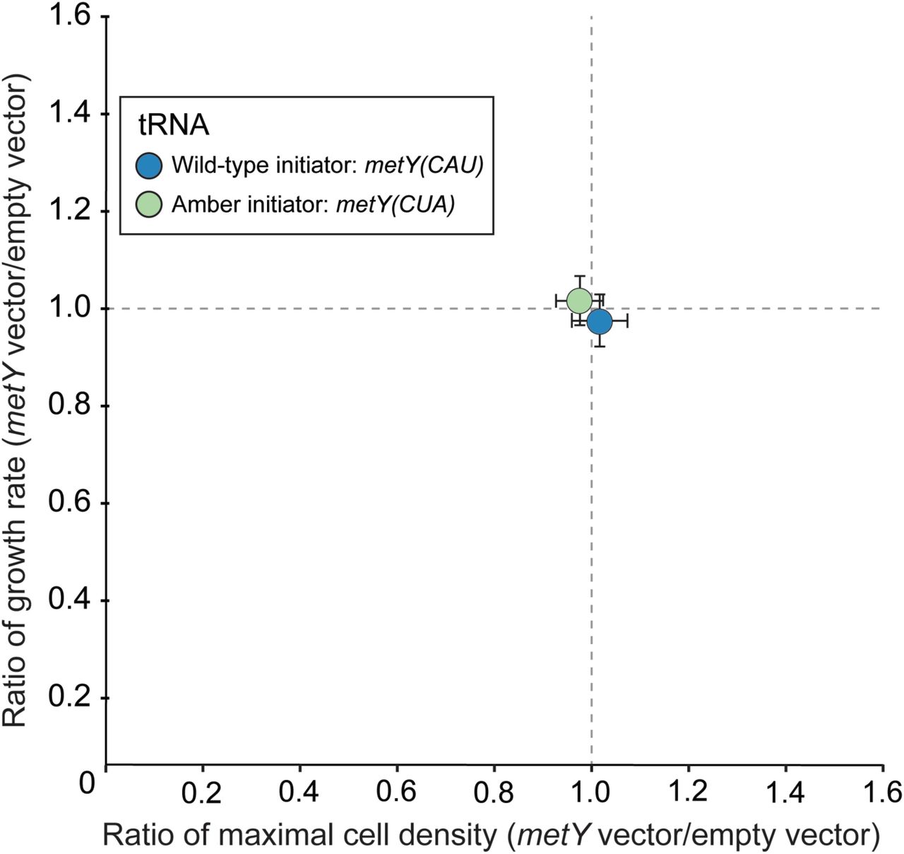
We next performed growth assays to measure the fitness of cells harbouring pULTRA::tac2metY(CUA). We compared growth characteristics of cells harbouring plasmids expressing the amber initiator tRNA (pULTRA::tac2metY(CUA)) and the wildY type initiator tRNA (pULTRA::tac2metY) to the empty vector (pULTRA::tacYEmpty) under inducing conditions. We found no significant effect (pYvalue > 0.5) from expression of either initiator tRNA on maximal growth rate or maximal cell density (Figure 2 and Table S2).

Amber initiator and wildYtype initiator tRNA expression results in minimal fitness impact on C321.ΔA.exp cells. Ratios of maximal cell densities (horizontal axis) and growth rate (vertical axis) were determined for strains containing either pULTRA::tac2metY(CUA) or pULTRA::tac2metY(CAU) plasmid versus their corresponding strains with the empty vector pULTRA::tac2Empty. Strains unaffected by initiator tRNA expression are expected to have ratios of 1. Strains exhibiting slower growth are below the horizontal gray line, and strains exhibiting lower maximum cell density are to the left of the vertical gray line. All strains were grown under the same induced (1 mM IPTG) condition. Each color represents the average of biological replicates (n = 3), and error bars show one standard deviation.
To better understand the global proteomic effect of expressing the amber initiator tRNA in C321.ΔA.exp, as well as to investigate possible causes for the observed increase in AUG reporter fluorescence (Figure 1), we next measured global protein production in these cells. We employed the dataYindependent acquisition method SWATHYMS (Sequential Window Acquisition of all Theoretical fragment ion Mass Spectra) which enables the collection of large, accurate, and reproducible quantitative proteomic datasets.16Y17 We collected data from E. coli C321.ΔA.exp samples with and without  expression three and five hours postYinduction.
We first interrogated the SWATHYMS data set with the aim of detecting canonical E. coli proteins with altered abundance under  expression. Summed fragment ion peak areas (area under the curve) of measured precursor ions (peptides) allowed us to compare protein abundance between E. coli C321.ΔA.exp expressing
  versus the control. Three hours postYinduction we identified only two differentially expressed proteins that reached our significance threshold (foldYchange >1.5, pYvalue <0.01). The lactose operon repressor protein (LacI) was upYregulated 4.9Yfold because it was constitutively expressed from the mediumYcopy pULTRA::tacYmetY(CUA) plasmid (Figure 3A, brown point and Table S3). The FhuE receptor protein was also upY regulated 3.7Yfold (Figure 3A, purple point). Together, these data show that at three hours postYinduction, the amber initiator
  does not appear to have a largeYscale effect on the C321.ΔA.exp proteome.

 expression causes delayed downregulation of ribosomal proteins in C321.ΔA.exp cells. (A) Volcano plot of 1,803 quantified proteins from E. coli C321.ΔA.exp cells at 3Yhours and 5Yhours after amber initiator
  induction. The orange points represent LacI and purple points represent FhuE. Vertical grey dashed lines represent fold change threshold: log2 ≥ 1 or ≤ −1 while the horizontal grey dashed line represents significance threshold: Y log10 pYvalue ≥ 2 equivalent to pYvalue ≤ 0.01. Green points represent significantly upYregulated proteins, red point represent significantly downY regulated, blue points represent unaltered expression, and grey points represent proteins that did not reach the significance threshold (pYvalue ≥ 0.01). (B) HeatYmap of ribosomal proteins quantified in C321.ΔA.exp control cells versus C321.ΔA.exp(pULTRA::tacYmetY(CUA)) cells expressing
  . The C321.ΔA.exp control and C321.ΔA.exp(pULTRA::tacYmetY(CUA)) strains were grown in LB in the presence of 1 mM IPTG and harvested at three and five hours postYinduction.
In contrast to three hours postYinduction, at five hours we identified 262 differentially expressed proteins (Figure 3A and Table S4). To assess the biological relevance of the differentially expressed proteins, we performed enrichment analysis employing the Protein ANalysis THrough Evolutionary Relationships (PANTHER) functional annotation tool.18 This analysis revealed that  expression had a significant impact on levels of proteins involved in the ribosome and translation process, leading to enriched biological process categories such as ribosomal biogenesis and assembly, and peptide metabolic pathways (Figure S2 and Table S5). Our data also showed that ribosomal proteins were abundant in both
  -expressing and control cells three hours postYinduction, but that they declined five hours postY induction in control cells. In contrast, when
  was expressed in the cell, ribosomal protein levels did not decline (Figure 3B and Figure S3, cluster 3).
In addition to the sustained production of the majority of ribosomal structural proteins five hours postYinduction, eight ribosomal RNA (rRNA) modification enzymes, two initiation factors (IF2 and IF3), and elongation factor 4 (LepA) were all significantly upYregulated 2.0 Y 9.1Yfold compared to control cells (Table S4). This expression profile was similar to ribosomal proteins that continued to be expressed at levels similar to three hours postYinduction while levels in control cells declined (Figure S3, cluster 3). Notably, LepA plays a direct role in ribosomal biogenesis19 and a simultaneous increase in observed rRNA modification proteins could suggest a rise in mature ribosome concentration. A recent study has linked ribosomal rRNA biogenesis to the conserved anticodon stem sequence of the initiator tRNA.20 Our results complement the findings of this previous study by showing that ribosomal protein is also highly upregulated in response to increased cellular levels of initiator tRNA. We speculate that an increased number of mature ribosomes, in addition to upYregulated initiation factors (IF2 and IF3), could explain the increased translation from the AUG start codon reporter we observed (Figure 1B, right blue bars), rather than  directly interacting with AUG start codons.
Previous evidence showed that  is a poor substrate for aminoacylYtRNA synthetases13Y14, 21 and increases the level of uncharged tRNA in the cell21 triggering the tRNA catabolic pathways (Figure 4). Additionally, the levels of uncharged
  may arise due to peptidyl tRNA hydrolase activity on amber initiator tRNA charged with glutamine.22 This potential imbalance in charged to uncharged tRNA ratio can be seen in our experiment through the increased abundance of poly(A) polymerase (PAP I) and polynucleotide phosphorylase (PNPase), both involved in tRNA degradation.23

Proposed model of cellular effects of amber initiator  expression. Increased RNA polymerase subunits transcribe
  from pULTRA::tacYmetY(CUA) resulting in reduction of charged
  . Black arrows represent differentially regulated pathways, green text represent upYregulated proteins, and gray boxes depict unaltered pathways.
We also observe an upregulation of guanosine pyrophosphatase (RelA)24 that is a key player in the cellular response to amino acid starvation through the stringent response.25 Despite RelA upregulation, it is unlikely that cells expressing the amber initiator tRNA are activating the stringent response, which is normally characterized by a reduction in the number of ribosomal proteins and expression of starvation sigma factors.26 Neither of these responses are observed in our experiment, and instead we see sigma factor 70 (σ70) increased 2.9Yfold and increased ribosomal protein expression (Table S4). We do observe the upYregulation of leucineYresponsive regulatory protein (Lrp) and transcriptional regulator TyrR, normally induced during the stringent response.25 Both Lrp and TyrR, in the absence of leucine and aromatic amino acids respectively, induce amino acid biosynthesis.27Y28 Providing further evidence that the stringent response is not being activated in response to the amber initiator tRNA expression, the increased levels of transcriptional regulators Lrp and TyrR in our work did not result in upYregulation of associated amino acid biosynthesis pathways normally observed in the stringent response.
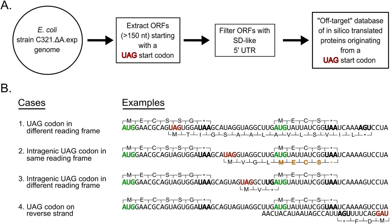
We next searched our SWATH dataset for offYtarget peptides produced from open reading frames (ORFs) beginning with UAG start codons in the C321.ΔA.exp genome. The C321.ΔA.exp strain has 321 instances of functional UAG stop codons in the genome replaced with UAA stop codons12 but we hypothesized that of the 27,108 UAG codons remaining in the genome, some may initiate translation in the presence of the amber initiator  . To identify peptides initiating from these UAG codons we generated an offYtarget proteome database using the genome sequence of C321.ΔA.exp (Figure 5A). We considered all genomic UAG codons as potential start codons and considered UGA and UAA as stop codons. We did not treat UAG as a stop codon because C321.ΔA.exp lacks release factor 1 which is required for termination at UAG stop codons. We generated a list of 27,108 ORFs and filtered out all ORFs shorter than 150 nucleotides (50 amino acids) in length since small proteins (less than 50 amino acids) are generally ignored in wholeYproteome studies to reduce background noise.29 This filtering process resulted in an offYtarget database of 9,857 ORFs initiating from UAG start codons. To further filter the ORF list we identified those ORFs with a ShineY Dalgarno (SD) sequence upstream of the UAG start codon, resulting in 990 ORFs in the final offYtarget database (Figure 5A and Table S6). Additionally, we considered that
  could be charged with glutamine in addition to methionine13Y14, 21 and thus created two versions of each protein in the database, one with an NYterminal methionine and one with a glutamine.

Method used to create UAG open reading frame offYtarget protein database. (A) Algorithmic workflow to create an offYtarget UAGYinitiating open reading frame (ORF) database from the C321.∆A.exp genome sequence. ORFs less than 150 nucleotides long were filtered out resulting in a reduction from 27,108 ORFs to 9,857. These ORFs were further filtered down to include only ORFs with ShineYDalgarnoYlike 5’ upstream sequences. The resulting 990 ORFs were then translated in two ways with UAG coding for methionine and glutamine to generate an offYtarget protein database. (B) UAG codon contexts that could result in ORFs with UAG start codon. The top proteins represent annotated sequences translated from canonical AUG start codon and bottom proteins represent hypothetical offYtarget protein sequences translated from a UAG start codon. ORFs displayed are for illustrative purposes only and are intentionally shorter than the minimum 150 nucleotide filter used to generate the offYtarget protein database. Green codons represent a canonical start codon; bold black codons represent a canonical stop codon in E. coli C321.∆A.exp; red codons represent a UAG start codon; brown amino acids represent shared peptides with sequences translated from a canonical AUG start codon and an amber UAG start codon.
We then combined the offYtarget database with the conventional E. coli MG1655 database (4,443 proteins) and searched our generated spectral library against it. Using the SWATHYMS data, we mined the combined spectral library and quantified 1,803 proteins across all three biological replicates that satisfied our significance criteria (protein and peptide level FDR <1% with at least 1 unique peptide observed). We observed 76 peptides that matched proteins from the offYtarget database, however, the identified peptides were shared with existing canonical E. coli proteins due to the presence of a UAG start codon upstream and inYframe with a canonical start codon (Figure 5B, case 2). We considered these peptides to originate from the conventional E. coli proteome since none were unique to the offYtarget database (Table S7). Despite not detecting any peptides originating from offYtarget ORFs we were able to detect peptides from four known proteins that have been previously shown to be present at 1Y5 copies per cell in E. coli grown in LB: P02942 (Tsr protein), P04949 (FliC protein), P07017 (Tar protein), and P39332 (YjgH protein).30 Together, these results show that despite detecting and quantifying over 1,800 proteins across a large dynamic range, we saw no evidence for  expression leading to offYtarget translation.
To further probe the possibility of translation initiation from genomically encoded offYtarget ORFs with UAG start codons, we built a singleYcopy reporter plasmid to determine whether we could detect protein production at the low levels expected for these ORFs. We designed and built the bacterial artificial chromosome (BAC) plasmids pSMARTYBAC::pRhaYNanoLuc(UAG) and pSMARTYBAC::pRhaYNanoLuc(AUG) (Figure 6A) and transformed them it into cells already containing the pULTRA::tacYmetY(CUA) plasmid or with the empty vector pULTRA::tacYEmpty. We performed a luciferase assay on cells induced with varying concentrations of rhamnose designed to induce varying amounts of NanoLuc luciferase production with constant induction of the amber initiator  or empty vector. As expected, we found that NanoLuc(UAG) luminescence was absolutely dependent on the amber initiator
  expression (Figure 6B).

SingleYcopy bacterial artificial chromosome (BAC) reporter plasmid expressing NanoLuc shows NYterminal amino acid is methionine and that peptides are only observed when the amber initiator tRNA is coYexpressed with the reporter. (A) SingleYcopy BAC NanoLuc plasmid design. Red ball and sticks represent transcriptional terminators; ori2, repE, and IncC encode singleYcopy origin. (B) NanoLuc luciferase reporter expression from C321.ΔA.exp cells harbouring either pULTRA::tacYEmpty or pULTRA::tacYmetY(CUA) tRNA expression plasmids along with either pSMART::pRhaY Nanoluc(UAG) or pSMART::pRhaYNanoluc(AUG) reporter plasmids. (C) Amino acid sequence of NanoLuc protein with peptides observed by PRM mass spectrometry highlighted in color. (D) Mass spectrometric identification of NYterminal amino acid of purified NanoLuc protein expressed with UAG start codon. Product ions specific to a methionyl NYterminal peptide from NanoLuc(UAG) shown with identical retention times and expected masses. Peptide sequence and preYcursor peptide fragmentation patterns shown above chromatogram. NanoLucY6xHis was NiYNTA purified from C321.ΔA.exp(pULTRA::tacYmetY(CUA); pSMART::pRhaYNanoLuc(UAG)) cells induced with 1 mM IPTG and 1 mM rhamnose. (E) NanoLuc peptides expressed from singleYcopy BAC detected in C321.ΔA.exp cells only when expressing the amber initiator. NanoLuc peptides observed in PRM mass spectrometry assay. Left, C321.ΔA.exp(pULTRA::tacY metY(CUA); pSMART::pRhaYNanoLuc(UAG)) cells; Right, C321.ΔA.exp(pULTRA::tacY Empty; pSMART::pRhaYNanoLuc(UAG)) cells.
At all rhamnose concentrations, when the amber initiator  was expressed, the luminescence resulting from NanoLuc(AUG) and NanoLuc(UAG) expression was equal (Figure 6B). We also found that the NanoLuc(AUG) reporter luminescence was independent of the presence of the amber initiator, having the same signal in the pULTRA::tacYEmpty cells as those containing pULTRA::tacYmetY(CUA).
This result is in contrast to what we found with the sfGFP reporter on a mediumY copy vector (Figure 1) where the presence of the amber initiator seemed to boost production of sfGFP(AUG). One explanation for this different behaviour could be that it is a result of the vector copy number: at lowYcopy number the mRNA encoding the NanoLuc protein is likely limiting production of the protein, whereas at much higher copy number the ribosomal abundance may be limiting sfGFP production. If this is true, and the expression of the amber initiator tRNA increases ribosomal abundance (Figure 3), then it follows that increased ribosome abundance in the medium copy sfGFP(AUG) reporter strain would cause an increase in fluorescence due to increased translation initiation from the reporter mRNA. In contrast, at lowYcopy increased ribosomal abundance has no effect on the NanoLuc(AUG) reporter because mRNA levels are limiting, not ribosomal abundance.
To identify the NYterminal amino acid incorporated into the NanoLuc reporter protein we purified the protein from C321.ΔA.exp cells harbouring the pSMARTY BAC::pRhaYNanoLuc(UAG) and pULTRA::tacYmetY(CUA) plasmids. We designed the pSMARTYBAC::pRhaYNanoLuc(UAG/AUG) plasmids to contain a CYterminal 6xHisYtag to enable purification of the fullYlength protein (Figure 6A). The internal and NYterminal peptides of the purified NanoLuc proteins were subjected to a targeted mass spectrometry technique called parallel reaction monitoring (PRM). The strength of the PRM method is that it provides high specificity and sensitivity and would allow us to specify peptides to target for accurate identification.31
The PRM assay showed that we could detect three peptides produced from tryptic digests of NanoLuc(UAG) (Figure 6C). The NanoLuc(UAG) NYterminal peptides contained methionine exclusively at the NYterminus (Figure 6D). The PRM assay was setup to agnostically target NYterminal peptides from NanoLuc(UAG) with any amino acid in the NYterminal position.22 This result is in contrast to previous work with the amber initiator tRNA, which showed both methionine and glutamine as the NYterminal amino acid from a reporter containing UAG as the start codon.32 However, these NY terminal amino acid differences could be a result of the different reporter gene used (NanoLuc versus CAT) or by the different bacterial strains used in the experiments (E. coli versus Mycobacterium smegmatis).
Using the PRM method and the singleYcopy NanoLuc(UAG) reporter plasmid, we searched for offYtarget peptides in C321.ΔA.exp cells harbouring the pSMARTY BAC::pRhaYNanoLuc(UAG) and pULTRA::tacYmetY(CUA) plasmids. We also measured peptides from cells containing the pSMARTYBAC::pRhaYNanoLuc(UAG) and pULTRA::tac2Empty plasmids as a control. In addition to inducing the pULTRA plasmid, we also weakly induced the pSMARTYBAC::pRhaYNanoLuc(UAG) reporter with 100 µM rhamnose (Figure 6B) as an internal control to determine our ability to detect UAGY initiating proteins from singleYcopy genes. Since the PRM method derives its sensitivity from limiting the peptides the mass spectrometer measures to a preYspecified list, it was not possible to target all 990 UAGYinitiating ORFs with a ShineYDalgarno sequence in C321.ΔA.exp. Instead, we limited our search space to peptides from 194 offYtarget ORFs with the strongest upstream ShineYDalgarno sequences (Table S6), in addition to the peptides of NanoLuc(UAG). Simultaneously, we targeted four weakly expressed E. coli host proteins previously observed in our SWATHYMS dataset. As before, we found no evidence for offYtarget protein expression from any of the offYtarget ORFs. We did detect one peptide that could have originated from a UAGYinitiating ORF, but this peptide was observed in both the empty vector control and amber initiatorYcontaining cells (Figure S5) suggesting that expression of this peptide is not dependent on  . At the same time, we were able to detect peptides from the four weakly expressed proteins from the E. coli host proteome (Table S11) along with NanoLuc(UAG) reporter protein from the singleYcopy pSMART plasmid, but only when cells contained pULTRA::tacYmetY(CUA), not pULTRA::tacYEmpty (Figure 6E).
In conclusion, we found that the amber initiator  efficiently initiates at the UAG amber stop codon from two different reporters, but does not initiate at detectable levels from genomic UAG codons. The amber initiator is thus shown to be orthogonal from a translation initiation point of view. Additionally, we showed that expressed NanoLuc(UAG) reporter proteins contain exclusively methionine at the NY termini, ensuring proteins created from this alternative initiation system have the same properties as wildYtype proteins. We also show for the first time that amber initiator expression causes increased ribosome levels, tRNA degradation, and upregulation of amino acid biosynthetic proteins, although these effects do not seem to affect strain fitness. Together, these results point towards a potential role for the amber initiator tRNA, after additional engineering to reduce its reliance on host aminoacylYsynthetases, in a fully orthogonal translation initiation system.33
METHODS
Bacterial strains
All designed and constructed plasmids were expressed in the genetically recoded organism E. coli strain C321.∆A.exp, which was a gift from George Church (Addgene #49018). All strain variants were grown in lysogeny broth Lennox (LBL) or on LBL agar, supplemented with zeocin for all E. coli strain C321.∆A.exp cultures and carbenicillin for cultures with reporter plasmid and spectinomycin for cultures with amber initiator plasmid.
Plasmid construction
pQEY60::T5YsfGFP reporter plasmids were designed to contain superfolder GFP (sfGFP) with AUG/UAG/GCC start codons. The pULTRA initiator tRNAY expression plasmids were created by using Gibson assembly34 to insert a 1971bp gBlock (IDT) contained metY genes from MG1655 (GenBank: U00096.3) with anticodon sequences of CAU (wildYtype initiator) and CUA (amber initiator) (Tables S8 and S9) into the pULTRAYCNF35 vector (Addgene #48215) (Figure 1A). To create an empty vector, we constructed the pULTRA::tacYEmpty plasmid via PCR backbone amplification of pULTRA::tac2metY(CUA) and ligation, such that all elements of the pULTRA::tac2 metY(CUA) plasmid except the metY(CUA) gene were retained. The pSMARTYBAC::pRhaY NanoLuc(UAG) and pSMARTYBAC::pRhaYNanoLuc(AUG) plasmids were constructed by cutting pSMARTYBAC with NotI followed by Gibson assembly with gblocks encoding a rhamnoseYinducible promoter (pRha) followed by the NanoLuc genes36 with a CY terminal 6xYHis tag and either UAG or AUG start codon. All plasmids were purified and stored in 10 mM TrisYCl, pH 8.5. These plasmids were transformed into C321.ΔA.exp strains containing either pULTRA::tacYmetY(CUA) or pULTRA::tacYEmpty.
Culture growth conditions for assay measurements
Bacterial cultures were grown overnight at 37° C in LBL with appropriate antibiotics. After overnight growth, each culture was diluted 1:100 into 400 µL LBL in a 96Ywell deep well plate containing either 1 mM IPTG to induce, or 2% glucose (w/v) to repress, metY(CUA) expression. To control for the effects of the reagents used for repression and induction of the tacI promoter, we used the same repressed (2% glucose) and induced (1 mM IPTG) culture conditions for all plasmid combinations, regardless of whether they contained a plasmid with a tacIpromoter or not.
Fluorescence measurements
Measurements of fluorescence intensity from the amber initiator plasmid system was performed as before15 with the following modifications: OD600 was used to estimate culture density, followed by fluorescence measurement (ex=485 nm, em=520 nm, bandwidth = 9 nm, PHERAstar FSX). Cultures from the above experiment were also measured on Cytoflex S flow cytometer (FITC, 488 nm excitation laser, 525/40 nm emission bandYpass filter). 10,000 measured events were triggered on a side scattering threshold. Data was processed using FlowJo v10.
Fitness analysis
Strains of C321.∆A.exp containing the pULTRA::tacYmetY(CAU), pULTRA::tacYmetY(CUA), or pULTRA::tacYEmpty plasmid were grown overnight as described above. Cell densities were measured, and cells were passaged to a starting OD600 of 0.1 into fresh 200 µL LBL with appropriate antibiotics and 1 mM IPTG. Cultures were grown in a flatYbottom 96Ywell plate sealed with gasYpermeable seal at 37°C (300 rpm). Absorbance at OD600 over time was measured on a SPECTROstar NANO plate reader at 5Yminute intervals. Growth rate (µr) and maximal cell density (max OD600) was determined for each culture using the R package GrowthCurver37 represented in Table S2.
Nanoluciferase Assay
Measurements of luminescence intensity from the amber initiator plasmid system was performed as before15 using the NanoYGlo Luciferase Assay System kit (Promega, #N1110) with the following modifications: expression of nanoluciferase was induced with the following concentrations of rhamnose: 0, 5, 50, 100, 500 µM, while 100 μL of culture were mixed with 100 μL of assay buffer and incubated for 15 min at room temperature, followed by luminescence measurement (em=230Y750 nm, gain = 900, PHERAstar FSX).
NanoLuc expression and purification
Single colonies from C321.ΔA.exp strain harbouring pULTRA::tacYmetY(CUA) and pSMARTYBAC::pRhaYNanoLuc(TAG) or pSMARTYBAC::pRhaYNanoLuc(ATG) as a control was used to inoculate 3 mL of LBL supplemented with zeocin, spectinomycin, and chloramphenicol and shaken at 37°C overnight. We used the overnight cultures to purify CYterminal 6X histidineYtagged nanoluciferase protein following NiYNTA purification as before15 with the following modifications: expression of  and nanoluciferase was induced with 1 mM IPTG and 1 mM rhamnose.
Sample preparation for mass spectrometry proteome analysis
Strains were grown overnight and back diluted 1:100 into fresh media in 250 mL flasks with appropriate antibiotics and 1 mM IPTG. Cells were harvested three hours and five hours postY induction (3,500 x g/4°C/10 min). Cell pellets were lysed with 0.5 mL CelLytic™ B Cell Lysis buffer (SigmaYAldrich #B7310) adding 0.1 mg lysozyme, 1X protease inhibitor (Roche #04693132001), and 25 units of benzonase (SigmaYAldrich #E1014). The lysate was incubated (15 min/room temperature) then centrifuged (16,000 x g/room temperature/10 min). 1 mL of soluble protein supernatant was collected per culture and analysed with minor changes from previous work.38. Proteins from whole cell lysate and NiYNTA purified fractions were precipitated with 1 mL iceYcold acetone (Y 20°C/overnight) then centrifuged (15,000 x g/4°C/10 min), followed by protein pellet resuspension in 100 mM TrisYHCl, 8 M urea (pH 8.0). Protein samples were digested with 1:100 (w/w) protein/trypsin (37°C/overnight), then digested again (37°C/4 hours) with fresh trypsin as before.
Peptide library generation
To produce a peptide ion library, samples from biological triplicates were pooled and separated by highYpH RPYHPLC as described previously.39 Mass spectrometry analysis was performed on a TripleTOF 6600 in dataYdependent mode as previously described39 except selection of ion precursors were performed over 140Yminute runs. Spectra were interrogated using ProteinPilot 5.0 using the Paragon algorithm (SCIEX)17 and searched against the complete E. coli KY12 MG1655 protein sequence database from Uniprot (proteome IDYUP000000625 (last modified 2nd April, 2018)) and inYhouse generated C321.ΔA.exp UAG offYtarget database (File S5). The default paragon algorithm includes a fixed cysteine carbamidomethylation modification, caused by dithiothrietol and iodoacetamide treatment, and variable modifications sourced from http://unimod.org.16 Proteins and peptides were accepted with a protein false discovery rate of <1%.
SWATHMMS analysis
Spectra collection was performed in data independent mode as described previously39 except, MS/MS spectra were collected across the m/z range of 350Y1500 with a 35 ms accumulation time. Ion libraries of individual strains were imported into PeakView software 2.1 using the SWATH MicroApp 2.0 (SCIEX) and matched against SWATHYMS data of individual replicates. Retention time calibration was performed using endogenous peptides and data were processed as previously described39 to generate cumulative protein areas from extracted ion chromatograms which were exported for further analysis (Figure S3 and S4). Proteins with two or more peptides, pYvalue < 0.01, and foldYchange >2 and <0.5 were considered as significantly differentially expressed (Tables S3 and S4).
Gene ontology analysis
The differentially expressed proteins were analysed against the total set of 1,803 quantified proteins using the overrepresentation test in PANTHER (http://pantherdb.org) with default parameters.18 Gene ontology biological processes with p < 0.05 (after Binomial test with Bonferroni multiple hypothesis test correction) were considered significant.
Generation of inMhouse offMtarget database
To identify peptides initiating from these genomic UAG codons we generated an offYtarget proteome database using the genome sequence of C321.ΔA.exp. We considered all genomic UAG codons as potential start codons and considered UGA and UAA as stop codons. We did not treat UAG as a stop codon because C321.ΔA.exp lacks release factor 1 which is required for termination at UAG stop codons. We generated a list of 27,108 ORFs and filtered out all ORFs shorter than 150 nucleotides (50 amino acids) in length. This process resulted in an offYtarget database of 9,857 protein sequences that could potentially form functional ORFs initiating from UAG start codons (Figure 5A). To determine the SDYlike sequences upstream of potential ORFs, we used a previously described method40 to predict thermodynamic interactions between the antiYShine Dalgarno and the upstream sequences of the potential ORFs, using the RNA coFold method of the ViennaRNA Package 2.041. We used a similar binding energy threshold of Y4.5 kcal/mol to filter down the offYtarget database, resulting in 990 potential offYtarget proteins with upstream SDYlike sequences (Table S6).
Targeted protein mass spectrometry of NMterminal peptide and offMtarget proteins
The targeted mass spectrometry method of parallel reaction monitoring (PRM)31 was used to identify the NYterminal peptide species of the nanoluciferase reporter protein with a UAG start codon. Additionally, we analysed the whole cell proteome for offYtarget protein expression. Samples were analysed on a high resolution QYExactive mass spectrometer (ThermoFisher Scientific) coupled to an EASYYnLC1000 liquid chromatography system. Peptides were eluted over a 120Yminute linear gradient with increasing concentration of elution buffer (99.9% (v/v) acetonitrile, 0.1% formic acid). The analysis consisted of one survey (full) scan at 70,000 resolution (400 m/z) for targeted precursors (Table S10 and Table S11) (AGC target 2e5, maximum injection time 100 ms, 2 m/z isolation width). Measured precursor ions were selected for HCD fragmentation with normalized collision energy of 30 followed by full ms/ms of product ions at 17,500 resolution (m/z 200, AGC target of 2e5 and 60 ms maximum injection time). Precursor and fragment ion spectra were analysed using Skyline version 4.2. 42 PRM assay result for identification of NYterminal amino acid can be downloaded from the Panorama web repository server (https://panoramaweb.org/H5K69A.url).
SUPPORTING INFORMATION
Figure S1: Fold change in fluorescence due to  induction; Figure S2: Proportion of differentially expressed proteins represented in each gene ontology category; Figure S3: SWATH protein expression profile clusters; Figure S4: Evaluation of SWATH YMS quantification technique; Figure S5: OffYtarget peptide detected in C321.ΔA.exp cells regardless of amber initiator expression; Table S1: Translation initiation efficiency; Table S8: Reporter and tRNA gene sequences; Table S9: Synthesized oligonucleotides (PDF).
Table S2: A) Growth curve data represented as OD600 measured every 5 minutes B)Average specific growth rate (minY1) and maximal OD600 (A.U.) (XLSX); Table S3: List of all quantified proteins using SWATHYMS technique three hours postYinduction; Table S4: List of all quantified proteins using SWATHYMS technique five hours postYinduction; Table S5: Gene ontology categorization of differentially expressed proteins five hours postYinduction; Table S6: List of offYtarget ORFs harbouring SDYlike sequences upstream; Table S7: List of all offYtarget proteins harmonized with Uniprot IDs from conventional E. coli database; Table S10: Parallel reaction monitoring targeted precursor list for NYterminal amino acid identification; Table S11: Parallel reaction monitoring targeted precursor list for offYtarget peptide identification (XLSX).
Supporting File S1: Amber initiator expression plasmid pULTRA_tacYmetY(CUA) (GB); Supporting File S2: Empty vector control plasmid pULTRA_tacYEmpty (GB); Supporting File S3: GFP reporter plasmid pQEY60_T5YsfGFP(UAG) (GB); Supporting File S4: Nanoluciferase reporter plasmid pSMARTYBAC_pRhaYNanoluc(UAG) (GB); Supporting File S5: FASTA file of combined proteomic database including potential offYtarget proteins for mass spectrometry identification (FASTA).
AUTHOR INFORMATION
Author Contributions
RMV and PRJ conceived the study. RMV and BWW performed experiments, analyzed data, and created figures and tables. RMV and PRJ drafted the manuscript. All authors read and approved the final manuscript.
Conflicts of Interest
The authors declare no competing conflicts of interest.
ACKNOWLEDGEMENTS
We thank Matthew McKay, Ardeshir Amirkhani, David Cantor, Karthik Kamath and Dana Pascovici of the Australian Proteome Analysis Facility for assistance in protein and data analysis. We also thank Michael A. Sorensen, Richard Fahlman, Olivier Borkowski, Heinrich Kroukamp, and Jeff Glasgow for helpful discussions. PRJ was supported by the Molecular Sciences Department, Faculty of Science & Engineering, and the Deputy ViceY Chancellor (Research) of Macquarie University. BWW and RMV are recipients of the Macquarie University Research Excellence PhD scholarship (MQRES). Aspects of this research was conducted at the Australian Proteome Analysis Facility, facilitated by the Australian Government’s National Collaborative Research Infrastructure Scheme.
ABBREVIATIONS

- amber initiator tRNA with CUA anticodon

- SWATHYMS
- Sequential Window Acquisition of all Theoretical fragment ion Mass Spectra
- IF1
- Initiation Factor 1
- IF2
- Initiation Factor 2
- IF3
- Initiation Factor 3
- sfGFP(AUG)
- superfolder green fluorescence protein gene with wildYtype AUG start codon
- sfGFP(UAG)
- superfolder green fluorescence protein gene with amber stop codon UAG as start codon
- NanoLuc(AUG)
- the NanoLuc gene with wildYtype AUG start codon
- NanoLuc(UAG)
- the NanoLuc gene with amber stop codon UAG as start codon


