

ABSTRACT
Aims Bacterial genotyping on the basis of the CRISPR array has been established in Mycobacterium tuberculosis with a method called spacer oligonucleotide typing (spoligotyping). The spoligotyping method had been widely used for both detection and typing of M. tuberculosis complex bacteria. This present study aimed at determining if the CRISPR array in Salmonella spp. could be applied to establish a correlationship between serogroup and the fingerprint generated by CRISPR typing.
Methodology and results A total of 30 samples were obtained from Diagnostic Veterinary Laboratory, Kota Kinabalu, Sabah. Serogroup was determined on the basis of ELISA (enzyme-linked immunosorbent assay). Four different serogroups were identified which were serogroup B, C, D, and E. DNA (deoxyribonucleic acid) was extracted and PCR (polymerase chain reaction) was performed using primers which were designed to amplify the CRISPR array in Salmonella genome. Our results indicate that there is a correlationship between serogroup obtained using ELISA and the profile generated by CRISPR typing.
Conclusion, significance and impact of study CRISPR typing has the potential to be applied for the genotyping of Salmonella.
INTRODUCTION
The first CRISPRs locus were identified over 25 years ago in Escherichia coli as ambiguous repeat (Ishino et al., 1987) and are known as CRISPR spacer arrays now (Mojica et al., 2000; Jansen et al., 2002a; 2002b). CRISPR arrays consists of tandem direct repeats (DRs) of 23 to 55 bp (base pair) in length interspaced by equal sized of variable spacer sequences that acquired from bacteriophages or plasmids (Bolotin et al., 2005; Mojica et al., 2005; Pourcel et al., 2005; Boyaval et al., 2007). The spacer in CRISPR locus was first applied to subtyping Mycobacterium tuberculosis strains and this method was known as spacer-oligonucleotide typing or “spoligotyping” (Groenen et al., 1993; Kamerbeek et al., 1997). Recently, the “next generation” microbead-spoligotyping method was applied to Salmonella in an assay named CRISPOL (for “CRISPR polymorphism”)(Fabre et al., 2012). Fabre et al. (2012) state that there are at least three potential interest of using the polymorphism in Salmonella CRISPR locus for clinical microbiology or public health laboratories which are: 1. CRISPR sizing by PCR to compare different isolate of Salmonella spp. 2. CRISPOL assay to subtype Salmonella serotype Typhimurium or its monophasic variant. 3. Development of PCR assay to target specific Salmonella serotype or strain. We aimed to demonstrate that CRISPR based genotyping can be utilized as a diagnostic tool to differentiate different Salmonella serogroups prevalent in Sabah.
MATERIAL AND METHODS
Sample Collection
Salmonella samples (N=30) were obtained from the Diagnostic Veterinary Laboratory, Kota Kinabalu, Sabah. All the Salmonella samples were isolated from avian host. The Salmonella bacteria were cultured on nutrient agar and MacConkey agar. The MacConkey agar is a selective medium for Salmonella which functions to ensure that contaminants are eliminated. The Salmonella colony from nutrient agar were transferred to nutrient broth and cultured overnight in incubator shaker at 37°C and 200 rpm (revolutions per minute). The Salmonella samples were stored at 25% glycerol stock in −80°C freezer for future use.
Serogrouping
ELISA was carried out as follows. Salmonella were cultured overnight at nutrient agar by streak plate method at 37°C. 0.85% saline solution was used for the ELISA test. The ELISA test was done using the Salmonella Sero-Quick Group Kit from Statens Serum Institut, Denmark. The protocol for Salmonella ELISA test was done according to the manufacturer protocol.
DNA Extraction
DNA was extracted according to modified Kang et al. (1998) method. The dry DNA pellet was re-suspended in 100 µl of 1X TE (Tris-EDTA) buffer and stored in −20°C for future use.
PCR of invA gene
The primer pair used for this PCR were taken from Cortez et al. (2006). PCR was performed in a reaction volume of 25 µl using the GE Healthcare illustraTM puReTaq Ready-To-Go PCR Beads. The preparation of PCR master mix was prepared according to manufacturer protocol. Amplification was carried out in a thermal cycler (MJ Research PTC-200 Peltier Thermal Cycler) using 35 cycles consisting of denaturation for 30 sec at 94°C, annealing for 1 min at 55°C, and extension for 1 min at 72°C, followed by a final extension for 7 min at 72°C. Electrophoresis of amplified products was carried out using 1.0% agarose gel in 1X TBE (Tris/Borate/EDTA) running buffer. The amplified DNA fragments were stained with ethidium bromide and visualized under UV (ultraviolet) light. A 100 bp DNA ladder (New England Biolabs Quick-Load 100 bp DNA Ladder) was used as a reference standard.
Primer Design
Design of CRISPR specific primers was carried out as follows. The complete genome of Salmonella enterica subsp. enterica serovar Typhi strain CT18 was first retrieved from the NCBI (National Centre for Biotechnology Information) with accession number of NC_003198.1. The genome was then analyzed for presence of CRISPR locus using online web tool called CRISPERFinder at http://crispr.upsud.fr/Server/CRISPRfinder.php. Once the CRISPR locus sequence was identified three pairs of primer set were designs to amplify the CRISPR 1 and CRISPR 2 locus presence in Salmonella genome using online web tool called Primer3 at http://bioinfo.ut.ee/primer3-0.4.0/. The primer sequences are listed in Table 1.0.
List of primer used for the CRISPR typing.
PCR of CRISPR Locus
PCR was performed in a reaction volume of 20 µl containing 1X Dream Taq. Green Buffer (Thermo Scientific), 0.1 mM dNTPs (deoxynucleoside triphosphate), 2 mM MgCl2 (Magnesium chloride), 5 µM of forward and reverse primer, 1 Unit of Thermo Scientific Dream Taq DNA polymerase and 1 µl of DNA template. Amplification was carried out in a thermal cycler (MJ Research PTC-200 Peltier Thermal Cycler) with initial denaturation of 96°C for 4 min, followed by 35 cycles of 96°C for 30 sec, 51°C for 30 sec, 72°C for 1 min and final extension step at 72°C for 2 min. All steps were the same for all primers except for the annealing temperature. The annealing temperature for CRISPR1 primer pair is 48°C, 51°C for CRISPR2 primer pair and 49°C for FARH primer pair. Electrophoresis of amplified products was carried out using 1.0% agarose gel in 1X TBE running buffer. The amplified DNA fragments were stained with ethidium bromide and visualized under UV light. A 100 bp DNA ladder (New England Biolabs Quick-Load 100 bp DNA Ladder) was used as a reference standard.
RESULTS
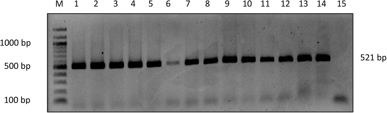
Salmonella samples (N=30) were test by ELISA for serogrouping. 53% of the samples were from serogroup C, followed by serogroup E with 20%, 17% for serogroup B and 10% for serogroup D. The genus confirmations for all the samples were done by the PCR of invA gene (Figure 1.0, 2.0 and 3.0). The expected size for the PCR product is 521 bp. PCR to amplify the CRISPR locus on Salmonella genome were done to differentiate the different Salmonella serogroups (Figure 4.0, 5.0 and 6.0).

PCR was done to amplify the invA gene on the Salmonella bacteria genome for genus confirmation. Lane M: 100 bp marker, lane 1 to 14: The PCR product of Salmonella invA gene and lane 15: Negative control without DNA template.

PCR was done to amplify the invA gene on the Salmonella bacteria genome for genus confirmation. Lane M: 100 bp marker, lane 1 to 9: The PCR product of Salmonella invA gene and lane 10: Negative control without DNA template.

PCR was done to amplify the invA gene on the Salmonella bacteria genome for genus confirmation. Lane M: 100 bp marker, lane 1 to 11: The PCR product of Salmonella invA gene and lane 12: Negative control without DNA template.

CRISPR1 primer pair was designed for PCR to amplify the CRISPR locus in Salmonella genome for serogrouping differentiation by molecular method. The above agarose gel picture show the amplification profile when Salmonella genome was tested with the designed primer. Lane M: 100 bp marker, lane1-3: Salmonella from serogroup B, lane 4-6: Salmonella from serogroup C, lane 7-9: Salmonella from serogroup D, lane 10-12: Salmonella from serogroup E and lane 13: Negative control without DNA template.

CRISPR2 primer pair was designed for PCR to amplify the CRISPR locus in Salmonella genome for serogrouping differentiation by molecular method. The above agarose gel picture show the amplification profile when Salmonella genome was tested with the designed primer. Lane M: 100 bp marker, lane1-3: Salmonella from serogroup B, lane 4-6: Salmonella from serogroup C, lane 7-9: Salmonella from serogroup D, lane 10-12: Salmonella from serogroup E and lane 13: Negative control without DNA template.

FARH primer pair was designed for PCR to amplify the CRISPR locus in Salmonella genome for serogrouping differentiation by molecular method. The above agarose gel picture show the amplification profile when Salmonella genome was tested with the designed primer. Lane M: 100 bp marker, lane1-3: Salmonella from serogroup B, lane 4-6: Salmonella from serogroup C, lane 7-9: Salmonella from serogroup D, lane 10-12: Salmonella from serogroup E and lane 13: Negative control without DNA template.
DISCUSSION
The O antigen present in the cell surface of Salmonella is extremely polymorphic and is used to determine the bacteria serogroup (Bee & Kwai, 2009). The variation in O antigen structure is due to the different types of sugar present, the arrangement of sugars, the addition of branch sugars and the modifying side groups in which such variation is used to serogroup Salmonella isolates (Wyk & Reeves, 1989; Fitzgerald et al., 2003; Luk et al., 2006). In this study four serogroups were found which were serogroup B, C, D and E. This finding is similar with the finding of Lindberg & Le Minor (1984) and Luk & Lindberg (1991) that stated over than 95% of the Salmonella strains that cause infection in human and animal is originate from the serogroup A to E. The identity of the Salmonella samples recruited for this study was confirmed by the present of invA gene in Salmonella genome using PCR method. This was done in order to ensure that all the samples used in this study were from the pure culture of Salmonella bacteria. PCR was an effective, rapid, reliable and sensitive method for the detection of invA gene present in Salmonella genome (Zahraei-Salehi et al., 2006). A study from Galán & Curtiss (1989) show that a group of genes (invA, B, C, D) confer Salmonella the ability to invade cultured epithelial cells. The use of this gene for Salmonella identification by PCR method has recently been suggested as this gene were shown to be found in a number of Salmonella strain (Galán & Curtiss, 1991). A study by Zahraei-Salehi et al. (2006) confirm that the invA gene sequence is unique to Salmonella and this unique sequence can be used as the PCR target to differentiate Salmonella from other organisms. The CRISPR typing was done to differentiate Salmonella serogroup by molecular method and the result was compared with the traditional serogrouping by ELISA method. The amplification profile of Salmonella from different serogroups were observed and compared. The principle of this method is that the variation in spacer number that interspaced between the direct repeats will give different lengths of CRISPR array and can be used to rapidly screen Salmonella isolates by PCR and gel electrophoresis analysis (Shariat & Dudley, 2014). The CRISPR1 and CRISPR2 primer pairs failed to differentiate some of the samples when compared to the serogrouping result obtained by the ELISA method. Depending on the antibody use for ELISA test, they may lack specificity because of the non-specific agglutination that might happen in some Salmonella bacteria as stated by Cheesbrough & Donnelly (1996). Only the CRISPR primer designated as FARH primer was able to completely differentiate all the samples from different serogroups. The reason why CRISPR1 and CRISPR2 primer cannot resolve the serogroup while FARH primer can might be because of the location of the primer along the CRISPR gene. The positions of forward and reverse primer for CRISPR1 along the gene are at nucleotide number 2,925,620 to 2,925,640 and 2,926,778 to 2,926,798 respectively. For CRISPR2 the positions of the forward and reverse primer along the gene are at nucleotide number 2,926,215 to 2,926,233 and 2,926,450 to 2,926,470 respectively. For the FARH the positions of forward and reverse primer along the gene are at nucleotide number 2,926,294 to 2,926,313 and 2,926,450 to 2,926,470 respectively. It can be seen from the results that some samples from the same serogroup have different amplification profile. This might be because of in one serogroup there were sub-group present as shown in White-Kauffmann-Le-Minor scheme (Grimont & Weill, 2008). Our findings are consistent with those obtained by Fabre et al. (2012) and Shariat & Dudley (2014) who used rapid CRISPR size typing to screen Salmonella spp. isolates by comparing the amplicon size of PCR product run on agarose gel electrophoresis.
CONCLUSION
From this study, it shown that only the FARH primer is able to resolve the samples into their respective serogroup. The serogrouping by ELISA method can be complement with CRISPR typing by PCR method to get more accurate result as non-specific agglutination might occur using the ELISA method.
ACKNOWLEDGEMENTS
We would like to thank UMS (Universiti Malaysia Sabah) for supporting this project under the UMSGreat grant with code project of GUG0106-1/2017. We also would like to thank the Veterinary Laboratory, Kota Kinabalu, Sabah for providing us the Salmonella samples used in this study.
Footnotes
Co-author: Normah Yusop, Diagnostic Veterinary Laboratory, 88999, Kota Kinabalu, Sabah, Malaysia, (h/p), (fax), Normah.Yusop{at}sabah.gov.my; Farhan Nazaie Nasib, Biotechnology Research Institute, Universiti Malaysia Sabah, 88400, Kota Kinabalu, Sabah, Malaysia, +6011-20701995 (h/p), farhannazaie12{at}gmail.com
REFERENCES


