

Abstract
Differential fluorescent labelling of bacteria has become instrumental for many aspects of microbiological research, such as the study of biofilm formation, bacterial individuality, evolution, and bacterial behaviour in complex environments. We designed a variety of plasmids, each bearing one of eight unique, constitutively expressed fluorescent protein genes in conjunction with one of four different antibiotic resistance combinations. The fluorophores mTagBFP2, mTurquoise2, sGFP2, mClover3, sYFP2, mOrange2, mScarlet-I, and mCardinal, encoding for blue, cyan, green, green-yellow, yellow, orange, red, and far-red fluorescent proteins, respectively, were combined with selectable markers conferring tetracycline, gentamicin, kanamycin, and/or chloramphenicol resistance. These constructs were cloned into three different plasmid backbones: a broad host-range plasmid, a Tn5 transposon delivery plasmid, and a Tn7 transposon delivery plasmid. The utility of the plasmids and transposons was tested in bacteria from the phyla Actinobacteria, Proteobacteria, and Bacteroidetes. We were able to tag representatives from the phylum Proteobacteria at least via our Tn5 transposon delivery system. The here constructed plasmids are available to the community and provide a valuable tool to investigate bacteria-bacteria, bacteria-host, and bacteria-environmental interactions.
Introduction
Labelling bacterial strains using fluorescent proteins have been used in manifold investigations, such as the study of biofilm formation, bacterial individuality and evolution, and bacterial behaviour in complex environments (e.g., plant surfaces, soil, or the mammalian gut) (Diard et al., 2013; Kroupitski et al., 2009; Monier and Lindow, 2005; Remus-Emsermann et al., 2013b; Remus-Emsermann and Schlechter, 2018; Tecon and Leveau, 2012; Tolker-Nielsen and Molin, 2000; Whitaker et al., 2017). Such studies require the equipment of bacteria with bright, stable, fast maturing, and highly abundant fluorescent proteins, which ideally offer unique spectral properties to unambiguously distinguish between differently labelled fluorescent bacteria. This is especially true during non-invasive in situ studies without additional staining procedures, such as fluorescence in situ hybridisation, and where autofluorescence and low signal-to-noise ratio might hinder observations (Remus-Emsermann and Schlechter, 2018).
To date, several mechanisms have been described by which fluorescent protein genes can be delivered into bacterial cells and/or integrated into their chromosomes (Andersen et al., 1998; Barbier and Heath Damron, 2016; Bloemberg et al., 2000; Choi and Schweizer, 2006; Lagendijk et al., 2010; Lambertsen et al., 2004; Ledermann et al., 2015; Miller et al., 2000; Remus-Emsermann et al., 2016a; Schada von Borzyskowski et al., 2015). Plasmids are attractive tools for fluorescent protein expression in bacteria, as they can be easily delivered into host cells and lead to high fluorescent protein production due to their presence in multiple copies (Million-Weaver et al., 2012). Plasmid stability usually requires a continuous selection (e.g., antibiotics (Andersson and Hughes, 2011)) and, while only some plasmids were found to be maintained in bacterial populations without the pressure exerted by antibiotics (Bloemberg et al., 2000; Rodriguez et al., 2017), the absence of a selective pressure was reported to lead to plasmid loss in growing bacterial populations (Lau et al., 2013; Smith and Bidochka, 1998; Summers, 1991). However, some experimental systems are not compatible with the use of antibiotics as a selective pressure. In natural environments, bacteria often occur as biofilms, where cells are fixed in space and enclosed in an extracellular polymeric substance matrix. Within a biofilm, individual cells experience heterogeneous environments, and owing to their location, they might either be exposed to or protected from antibiotic pressure (Stewart and Costerton, 2001).
To overcome the disadvantage of plasmids loss or their inability to replicate in some hosts, chromosomal insertion of fluorescent markers is advantageous. Chromosomal insertions cannot be lost in the same fashion as plasmids during division, and the probability of mutations that would disrupt the functionality of fluorescent protein genes is low. Several molecular tools and mechanisms exist to integrate chromosomal insertions into bacterial genomes, such as homologous recombination (Ledermann et al., 2015), CRISPR-Cas9 (Jinek et al., 2012), Zinc-finger nucleases (Carroll, 2011), or transposase-based systems (Liu et al., 2013; Peters, 2014; Reznikoff, 2008).
Transposon systems can differ widely in their host specificity and mode of integration. For instance, the Tn5 transposon has been shown to be functional in a wide range of Gram-negative bacteria and to randomly insert into their genomes with high efficiency (Reznikoff, 2008). This ability has been exploited to generate random mutations libraries (Christen et al., 2011; de Lorenzo et al., 1990), but also to integrate fluorescent protein genes into bacterial genomes (Andersen et al., 1998; Schada von Borzyskowski et al., 2015). In contrast to Tn5 transposons, Tn7 transposons only integrate into specific regions, such as the attTn7 site, in the host chromosome. Integration at attTn7 is mediated by four transposases, TnsA, B, C and D and appears to be prevalent in phylogenetically diverse species (Choi and Schweizer, 2006; McKenzie and Craig, 2006; Parks and Peters, 2007). Even though the host range of Tn7 transposons is limited compared to the Tn5 transposon, it has the tremendous advantage that the insertion does not disrupt any genes and has been argued to have little effect on bacterial fitness.
Here, we developed three sets of plasmids, which contain genes encoding for the latest generation of fluorescent proteins, i.e. that have blue, cyan, green, yellow, orange, red, and far-red fluorescence emissions, with different degrees of spectral overlap. The fluorescent protein genes mTagBFP2 (mTB2), mTurquoise2 (mTq2), sGFP2, mClover3 (mCl3), sYFP2, mOrange2 (mO2), mScarlet-I (mSc), and mCardinal (mCa) were combined with four combinations of antibiotic resistance genes, i.e. gentamicin, kanamycin, tetracycline and/or chloramphenicol, on three different plasmid backbones: a broad host-range plasmid, a Tn5 transposon delivery plasmid, and a Tn7 transposon delivery plasmid (Figure 1). The broad host-range plasmid contains the pBBR1 origin of replication (Miller et al., 2000). The Tn5 transposon plasmid pAG408 is based on an R6K origin of replication. It is a suicide plasmid that only replicates in presence of the π factor (Suarez et al., 1997). The Tn7 transposon plasmid pGRG36 is based on the thermo-unstable pSC101 origin of replication and is therefore a conditional suicide plasmid for species closely related to E. coli and a suicide plasmid in all other bacteria (McKenzie and Craig, 2006). The expression of the fluorescence protein genes is driven by the nptII promoter of the neomycin phosphotransferase gene (i.e. kanamycin resistance gene). The nptII promoter is considered constitutive, strong and was previously used to drive fluorescent protein expression from chromosomal insertions (Ledermann et al., 2015; Ramirez-Mata et al., 2018). We demonstrate the delivery of plasmids and transposon in a broad phylogenetic background of recently isolated environmental bacteria. Furthermore, we show the heterologous expression of fluorescent proteins at the single-cell resolution in those environmental bacterial strains using fluorescence microscopy.

Overview of cloning procedures. (A) Construction of the pMRE plasmid series. To obtain the pMRE13X plasmid series, HindIII-digested pFru97 was isothermally assembled with a fragment containing the ntpII promoter (PntpII) and a fragment carrying one of eight fluorescent protein genes (FP) - mTagBFP2, mTurquoise2, sGFP2, mClover3, sYFP2, mOrange2, mScarlet-I, and mCardinal. The rest of the pMRE series were constructed through the assembly of SacI-digested pMRE13X and one of three additional antibiotic resistance gene (AR) - Gentamicin (GentR), Kanamycin (KanR), and Tetracycline (TetR). (B) Construction of the transposon delivery plasmid series pMRE-Tn5 and pMRE-Tn7. PCR products using the pMRE plasmid series as templates containing AR, FP and a Chloramphenicol resistance (CamR) were isothermally inserted into EcoRV digested pAG408 to yield the pMRE-Tn5 series. Similarly, PCR products containing AR, FP and a Chloramphenicol resistance (CamR) were blunt-end cloned into SmaI digested pGRG36 to yield the pMRE-Tn7 series.
Materials and Methods
Media and growth conditions
Bacterial strains and their growth media are shown in Table 1. Growth media were prepared according to the manufacturer’s recommendations and supplemented with 1.5 % agar (Agar No.1, Oxoid) where commercial broth media was used as a base: lysogeny broth agar (LBA, Lysogeny broth Miller, Merck), tryptic soy broth agar (TSA, Merck), nutrient agar (NA, Nutrient broth, Oxoid), or Reasoner’s 2a agar (R2A, HiMedia). E. coli carrying pMRE or pMRE-Tn5 plasmids were cultivated at 37 °C, whilst pMRE-Tn7 plasmid-carrying cells were cultivated at 30 °C to prevent plasmid loss. All other strains used in this study were cultivated at 30 °C. For counterselection after conjugation, auxotroph E. coli S17-1 was grown on either MM2 agar medium (Zengerer et al., 2018) (4.0 g L-1 l-asparagine, 2.0 g L-1 K2HPO4, 0.2 g L-1 MgSO4 · 7H2O, 3.0 g L-1 NaCl, 10.0 g L-1 sorbitol, 15 g L-1 agar) or minimal agar medium (Harder et al., 1973) (1.62 g L-1 NH4Cl, 0.2 g L-1 MgSO4, 1.59 g L-1 K2HPO4, 1.8 g L-1 NaH2PO4 · 2H2O, 15 g L-1 agar, with the following trace elements: 15 mg L-1 Na2EDTA2·H2O, 4.5 mg L-1ZnSO4 · 7H2O, 3 mg L-1 CoCl2 · 6H2O, 0.6 mg L-1 MnCl2, 1 mg L-1 H3BO3, 3.0 mg L-1 CaCl2, 0.4 mg L-1 Na2MoO4·2H2O, 3 mg L-1 FeSO4·7H2O, and 0.3 mg L-1 CuSO4·5H2O) supplemented with 0.4 % w/v succinate or glucose were used, depending on the recipient strain (Table 1). Where appropriate, media were supplemented with antibiotics in the following concentrations: 100 mg L-1 ampicillin, 50 mg L-1 kanamycin, 20 mg L-1 gentamicin, 15 mg L-1 chloramphenicol, or 15 mg L-1 tetracycline.
Bacterial strains, their relevant naturally-occurring antibiotic resistances, standard growth media and conditions for selection after conjugation experiments.
Plasmid construction
All plasmids used or constructed in this study are shown in Table 2. Generic plasmid maps and cloning procedures are presented in Figure 1. For plasmid construction, PCRs were performed using Phusion High-Fidelity DNA polymerase (Thermo Scientific) following the manufacturer’s recommendations. Annealing temperatures (Ta) were chosen based on the respective melting temperature (tm) of the primers (Table 3). Touchdown PCRs were performed to amplify PCR products with overlapping ends for isothermal assemblies. To that end, the initial Ta was set to 10 °C above the lowest tm of the respective primers and the Ta was gradually reduced 1 °C per cycle for a total of ten cycles. After the tenth cycle, a 2-step PCR with Ta set to 72 °C was performed for 25 cycles.
Plasmids used in this work.
Primers used in this work.
Several plasmids that were used as source for fluorescent protein genes were acquired from the plasmid repository addgene.org: pGRG36 was a gift from Nancy Craig (Addgene plasmid #16666). pTriEx-RhoA-wt_mScarlet-I_SGFP2 (Addgene plasmid #85071) and pLifeAct-mTurquoise2 (Addgene plasmid #36201) were gifts from Dorus Gadella. pNCS-mClover3 was a gift from Michael Lin (Addgene plasmid #74236). Plasmids mCardinal-pBAD (Addgene plasmid #54800) and mTagBFP2-pBAD (Addgene plasmid #54572) were gifts from Michael Davidson and mOrange2-pBAD was a gift from Michael Davidson & Roger Tsien (Addgene plasmid #54531). pFru97 was a gift from Johan Leveau (Tecon and Leveau, 2012). All plasmids that were used for PCR amplification were isolated using the DNA-spin plasmid DNA purification kit (INtRON biotechnology) following the manufacturer’s recommendations. The designated plasmid backbones of the herein constructed plasmids, pFru97, pGRG36, and pAG408, were isolated using the Zyppy™ plasmid midiprep kit (Zymo) following the manufacturer’s recommendations. Gel purification and PCR clean-up was performed using the Monarch DNA Gel Extraction Kit (NEB) or the DNA Clean & Concentrator™-5 Kit (Zymo), respectively.
To construct the pMRE130 series, the promoter of the nptII gene (PnptII) was amplified from pRJPaph_gfp+ using primer FWD_PnptII, which contains overlapping sequences to HindIII-digested pFru97, and either primer REV_PnptII_1, REV_PnptII_2, or REV_PnptII_3, which contain overlapping sequences to the different fluorescent protein genes. The fluorescent protein genes were amplified using primer FWD_FP1, FWD_FP2, or FWD_FP3 with overlapping sequences to the PnptII fragment and primer X is a placeholder for either 0, 1, 2, 3, 4, 5, 6, or 7 representing mTagBFP2, mTurquoise, sGFP2, sYFP2, mOrange, mScarlet-I, mCardinal, or mClover3 encoding versions of the plasmids, respectively.
REV_FP1, REV_FP2, REV_FP3, REV_FP4, REV_FP5, or REV_FP6 with overlapping sequences to HindIII-digested pFru97. The two resulting fragments were mixed with the HindIII-digested pFru97 backbone and isothermally assembled to yield plasmids pMRE130, pMRE131, pMRE132, pMRE133, pMRE134, pMRE135, pMRE136, and pMRE137. To construct the pMRE140 plasmid series, the gentamicin resistance gene (GentR) was amplified from pAG408 using primers FWD_Gent and REV_Gent with overlaps to SacI-digested plasmids of the p MRE 13 X series, yielding pMRE140,pMRE141, pMRE142, pMRE143, pMRE144, pMRE145, pMRE146, and pMRE147. To construct the pMRE150 plasmid series, the kanamycin resistance gene (KanR) was amplified from pAG408 using primers FWD_Kan and REV_Kan with overlapping sequences to SacI-digested plasmids of the pMRE13X series, yielding pMRE150, pMRE151, pMRE152, pMRE153, pMRE154, pMRE155, pMRE156, and pMRE157. To construct the pMRE16X plasmid series, the tetracycline resistance genes (TetR) were amplified from pTE105-mChe using primers FWD_Tet and REV_Tet with overlapping sequences to SacI-digested plasmids of the pMRE13X series, yielding pMRE160, pMRE161, pMRE162, pMRE163, pMRE164, pMRE165, pMRE166, and pMRE167.
Isothermal assembly was performed as described previously (Benoit et al., 2016; Gibson et al., 2009; Remus-Emsermann et al., 2016a). In short, plasmid backbones and inserts with overlapping sequences were mixed in a 1:3 molar ratio (20-100 ng backbone) to reach a total volume of 5 µL. Then, 15 µL of isothermal assembly mix were added and the reaction was incubated for 15 minutes at 50 °C. Transformation of chemically competent E. coli strains was performed using standard procedures as recommended by the respective suppliers or as described in Sambrook et al. (Sambrook et al., 1989) (See Table 1).
To construct the pMRE-Tn5 plasmid series, pMRE1XX plasmids were used as a template to amplify the desired DNA fragments, which include one of the eight fluorescent protein gene and one of the four antibiotic resistance marker combinations. Cassettes were amplified as two fragments using the primers FWD1_pMRE_pAG408 and RV1_pMRE_pAG408 or FWD2_pMRE_pAG408 and RV2_pMRE_pAG408. Primers FWD1_pMRE_pAG408 and RV2_pMRE_pAG408 contain overlapping sequences to EcoRV-digested pAG408. For isothermal assemblies, EcoRV-digested pAG408 was mixed with the insert fragments in a 1:3:3 ratio as described above and assembled plasmids were transformed into chemically competent E. coli S17-1.
To construct the pMRE-Tn7 family, fluorescent protein genes and antibiotic resistance genes were amplified from the pMRE1XX series using primers FWD_pMRE-Tn7 and REV_pMRE-Tn7. The amplicons were gel purified using the Monarch DNA Gel Extraction Kit (NEB) and phosphorylated using T4 polynucleotide kinase (Life Technologies). pGRG36 was SmaI digested and dephosphorylated using thermosensitive alkaline phosphatase according to the manufacturers’ recommendations (Fast AP, Life Technologies). Following dephosphorylation, plasmids were purified using the DNA Clean & Concentrator™-5 Kit (Zymo). Amplicons were then cloned into linearised and dephosphorylated pGRG36 using Quick-Stick T4 Ligase (Bioline). Ligations were purified using the DNA Clean & Concentrator™-5 Kit and transformed into chemically competent E. coli (Top10, One Shot™ MAX Efficiency™ DH5α™-T1R, or Stellar).
All pMRE1XX series plasmids were verified by Sanger sequencing using primers FWD_seq and REV_seq at Macrogen. pMRE-Tn5-1XX and pMRE-Tn7-1XX series plasmids were verified by PvuII restriction digests at 37 °C for 1 h. To provide a convenient means of plasmid delivery, all plasmids were subsequently transformed into E. coli S17-1, which allows conjugations to recipient strains.
Conjugation
Recipients were grown on standard agar media for up to four days, depending on the growth rate of each environmental strain (see Table 1). Donor strains E. coli S17-1 were grown on LBA overnight (see Table 1). Plasmids delivered into recipient strains include pMRE135, pMRE145, pMRE165, pMRE-Tn5-143, pMRE-Tn5-145, pMRE-Tn5-165, pMRE-Tn7-145, or pMRE-Tn7-165. Freshly grown bacteria were harvested using a loop and resuspended in 1 × phosphate buffered saline (1 × PBS (8 g L-1 NaCl, 0.24 g L-1 KCl, 1.42 g L-1 Na2HPO4, 0.24 g L-1 KH2PO4)) to reach an OD600nm of 1. Each recipient strain was mixed with their respective donor strains in 1:1 ratio and the mix was then concentrated by centrifugation (4000 g, 5 min) to reach an estimated OD600nm of 20. Bacterial mixes were drop spotted on NA and incubated for 18 hours at 30 °C. For conjugations using pMRE-Tn7, 0.1 % w/v arabinose was added to the medium, as the Tn7 transposase genes are under the control of an arabinose-inducible promoter. After incubation, the cells were harvested and resuspended in 1 mL 1 × PBS. The bacterial mix was spread onto appropriate minimal agar media containing a sole carbon source (either 0.4 % w/v succinate or 0.4 % w/v glucose, see Table 1) and appropriate antibiotics. Depending on the recipient strain, transconjugants appeared within 5-10 days. To further counterselect against E. coli, single colonies were restreaked at least twice onto fresh minimal media before growing them on complex media.
Validation of transposon insertion events
To provide a convenient tool to assess successful insertions of Tn5 and Tn7 transposons, a multiplex PCR was designed to determine the presence of plasmid backbones and fluorescent protein genes in one reaction. Primers FWD_Tn5/7_gt and RV_Tn5/7_gt were designed to amplify the fluorescent protein genes for both Tn5 and Tn7 delivery systems, while the combinations FWD_Tn5_gt and RV_Tn5_gt and FWD_Tn7_gt, RV_Tn7_gt, and Tn7_gt amplify a specific fragment of the backbone of Tn5 and Tn7 plasmids, respectively. For Tn5 insertions, the primer mix FWD_tn5/7_gt, RV_tn5/7_gt, FWD_tn5_gt, and RV_tn5_gt was used. For Tn7 insertions, primer mix FWD_tn5/7_gt and RV_tn5/7_gt and primer mix FWD_tn7_gt, RV_tn7_gt, and Tn7_gt were used. Reactions were performed using the KAPA2G Fast HotStart ReadyMix PCR kit (Kapa Biosystems) following the manufacturer’s instructions.
Fluorescent protein absorption and emission spectra and fluorescence intensity
The absorption and emission spectra of the different proteins were measured in a Cary Eclipse Fluorescence Spectrophotometer (Agilent Technologies). To that end, overnight cultures of E. coli strains expressing the different fluorescent proteins were harvested by centrifugation (4000 g, 3 min), washed in 1 × PBS and resuspended to reach an OD600nm of 1. Then, 1 mL of each culture was measured in a polystyrene cuvette (VWR), besides cultures expressing mTagBFP2 and mCardinal, which were measured in a far-UV quartz cuvette (Agilent Technologies) due to their spectral properties. Absorption and emission were measured in the spectrophotometer in 1 nm intervals. The absorption and emission spectra of the proteins used in this study are available as “protein collection” on the webpage fpbase.org (https://www.fpbase.org/collection/78/; (Lambert, 2018)). This online platform allows for a convenient comparison of existing microscopy hardware and fluorophore properties.
To determine brightness of E. coli DH5α carrying plasmids or transposon insertions, respective strains were grown in eight biological replicates in LB broth (300 rpm, 37 °C) to reach an OD600nm of 1. Fluorescence intensity of cultures were measured in a flat bottom 96-well plate (Costar) using a FLUOstar Omega plate reader (BMG Labtech) equipped with an excitation filter (band pass 580/10) and an emission filter (band pass 620/10). Fluorescence intensity was corrected by background subtraction.
Fluorescence microscopy and image processing
Fluorescent protein-expressing E. coli were grown overnight in LB broth (210 rpm, 37 °C). Cultures were harvested by centrifugation (4000 g, 3 min) and washed in 1 × PBS. Bacterial cultures were resuspended in 1 × PBS to an OD600nm of 0.01, i.e. ∼107 bacteria/mL, and were mounted on agarose slabs (0.8 % w/v agarose in H2O). Fluorescence microscopy was performed on a Zeiss AxioImager.M1 fluorescent widefield microscope equipped with Zeiss filter sets 38HE, 43HE, 46HE, 47HE, and 49 (BP 470/40-FT 495-BP 525/50, BP 550/25-FT 570-BP 605/70, BP 500/25-FT 515-BP 535/30, BP 436/25-FT 455-BP 480/40, and G 365-FT 395-BP 445/50, respectively), an Axiocam 506 and the software Zeiss Zen 2.3. For confocal microscopy, a Leica SP5 confocal laser scanning microscope equipped with a 405 nm UV laser, an Argon laser line 458, 476, 514, nm, and a diode-pumped solid-state laser line 561 nm and software Leica LAS AF (version 2.6.3.8173) was used. Suspensions containing six or seven different fluorescent protein-tagged E. coli were detected through confocal microscopy using sequential scanning mode. For six colour mixtures consisting of mTB2, mTq2, mCl3, mO2, mSc, and mCa-expressing E. coli, a first scan using laser line 561 nm was used for emission windows of 570-599 nm, 600-630 nm, and 650-750 nm. Then, a second scan using laser lines 405, 458 and 476 nm were used for emission windows 440-460 nm, 480-500 nm, and 520-540 nm. For seven colour mixtures consisting of mTB2, mTq2, sGFP2, sYFP2, mO2, mSc, and mCa-expressing E. coli, a first scan using laser line 561 nm was used for emission windows of 570-599 nm, 600-630 nm, and 650-750 nm. A second scan using line 514 nm was used for the emission windows 540-560 nm. A third scan using lines 405, 458, 476 nm, and were used for the emission windows 440-460 nm, 480-500 nm, and 520-540 nm.
All image analysis and processing were carried out in ImageJ/FIJI (Schindelin et al., 2012). Signal bleed-through was corrected using a channel subtraction method. First, individual channels were thresholded (ImageJ/Fiji threshold using Otsu) to produce binary images. Binary template images were then dilated (ImageJ/Fiji command dilate) to accommodate spherical aberrations and subtracted from channels with signal bleed-through. All channels were background subtracted.
Single-cell fluorescence was analysed as described previously (Remus-Emsermann et al., 2016a). In short, bacteria were mounted on an agar slab as described above and samples were analysed using a Zeiss AxioImager.M1 at 100x magnification. Multichannel images were acquired using phase contrast and appropriate fluorescent filters (see above). Using ImageJ/Fiji, phase contrast channels were thresholded using standard settings and used as a mask to determine the mean fluorescence signal of individual particles in the respective fluorescent channels.
Results
In this work, a total of 96 plasmids were constructed (Table 2). The plasmids constitute three sets, including i) 32 broad-host range plasmids based on the pBBR1 origin of replication, ii) 32 suicide plasmids based on the R6K origin of replication, which are maintained solely in hosts carrying the π factor, and a Tn5-based transposon system, and iii) 32 narrow host-range plasmids harbouring a pSC101 temperature-sensitive origin of replication and the Tn7-based transposon system. Each set consists of plasmids that carry one of eight different fluorescent protein genes and one of four different combinations of antibiotic resistances. We used a simple naming convention and separated the plasmid into the pMRE13X-series, which all confer chloramphenicol resistance, the pMRE14X-series, which all confer gentamicin and chloramphenicol resistance, the pMRE15X-series, which all confer kanamycin and chloramphenicol resistance, and the pMRE16X-series, which all confer tetracycline and chloramphenicol resistance. The pFru97-based pMRE series contains an additional kanamycin resistance. Furthermore, we named plasmids based on the contained fluorescent protein, i.e. pMRE1X0 denotes mTagBFP2, pMRE1X1 denotes mTurquoise2, pMRE1X2 denotes sGFP2, pMRE1X3 denotes sYFP2, pMRE1X4 denotes mOrange2, pMRE1X5 denotes mScarlet-I, pMRE1X6 denotes mCardinal, pMRE1X7 denotes mClover3. Transposons are indicated as pMRE-Tn5-1XX for Tn5 transposons and pMRE-Tn7-1XX for Tn7 transposons.
Fluorescent protein-expressing bacteria were found to exhibit the expected fluorescence absorption and emission spectra (Figure 2) (Bindels et al., 2017; Chu et al., 2014; Goedhart et al., 2012; Kremers et al., 2007; Subach et al., 2011), suggesting that no changes in their spectral properties were introducing by cloning procedures. However, it was noticeable that bacterial cells harbouring the pMRE14X-series exhibited increased fluorescence intensities than in the rest of the plasmid series, likely due to readthrough of the gentamicin promoter which was cloned upstream the fluorescent protein gene. Therefore, the fluorescence intensity of the pMRE-series carrying the mScarlet-I gene in combination with all antibiotic resistance genes was determined in E. coli DH5α. Additionally, the emitted fluorescence was assessed in four independent E. coli DH5α::Tn5-MRE145 Tn5 insertions mutants and four independent E. coli DH5α::Tn7-MRE145 Tn7 insertion mutants. Plasmids pMRE135, pMRE155 and pMRE165 led to similar fluorescence in E. coli DH5α, however, plasmid pMRE145 resulted in significantly brighter fluorescence intensities (Figure 3A). After Tn5 and Tn7 transposition, E. coli DH5α exhibited similar fluorescence intensities, however, transposon-conferred fluorescence intensities were significantly lower than plasmid-conferred fluorescence intensities (Figure 3B).

Normalised (A) absorption and (B) emission spectra of the fluorescent proteins used in this work.

(A) Fluorescence intensity of E. coli DH5α cultures expressing mScarlet-I from different pMRE plasmid series (pMRE135, pMRE145, pMRE155, pMRE165). (B) Fluorescence intensity of mScarlet-I in plasmid-borne E. coli DH5α cultures (pMRE145), Tn5- and Tn7-insertion mutants. E. coli DH5α was used as control. One-way ANOVA and Tukey’s post hoc test were used to infer statistical differences between groups. Letters indicate differences among means with a 99.9% confidence interval (p < 0.0001) A.u.: arbitrary units.
Plasmid sets are broadly transmissible into a variety of Gram-negative bacteria
Plasmid functionality and fluorescence protein expression was tested in a phylogenetically broad range of bacteria that were recently isolated from plant leaves (Table 4). The pMRE-series plasmids were successfully conjugated and maintained in Sphingomonas melonis FR1, Erwinia amylovora CFBP1430S, Pantoea agglomerans 299R, and P. syringae B728a. Tn5 transposon plasmid were delivered by conjugation and integrated into the genome of Bradyrhizobium sp. Leaf396, Methylobacterium sp.
Bacterial strains used as recipients for conjugation of pMRE, pMRE-Tn5 and pMRE-Tn7.
Leaf92, S. melonis FR1, Sphingomonas phyllosphaerae FA2, E. amylovora CFBP1430S, P. agglomerans 299R, and Pseudomonas citronellolis P3B5. Tn7 transposon plasmids were delivered by conjugation and integrated into the genome of S. melonis FR1, E. amylovora CFBP1430S, and P. agglomerans 299R.
For a fast assessment of transposon-mediated insertions, multiplex PCRs were developed to amplify a region of the insert (i.e. fluorescent protein gene) and plasmid backbone simultaneously, i.e. a PCR on the donor plasmid resulted in two PCR products, while a genomic integration of the transposon resulted in only one PCR product. As expected, successfully integrated insertions into the genome of all tested strains were corroborated, as no plasmid backbone was detected whilst the fluorescent protein gene was correctly amplified (Supplementary Figure 1).
The provided fluorescent protein toolbox allows to track multiple bacterial strains in parallel
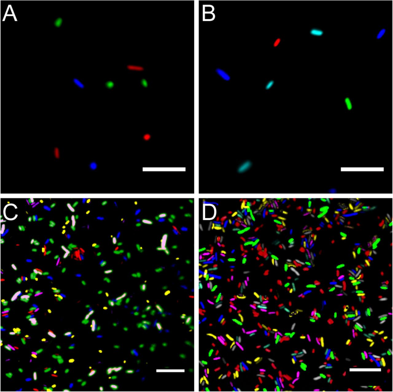
Using standard fluorescence microscopy equipment, it was possible to track up to seven bacterial strains in parallel. Employing widefield fluorescence microscopy and widely distributed fluorescence filter sets (i.e. standard blue, green and red fluorescence emission filters) three bacterial strains could be unambiguously identified. Thereby allowing to monitor three strains equipped with mTB2, mCl3, and mSc (Figure 4 A), respectively. Less common combination of filters (i.e. blue, cyan, yellow, and red fluorescence emission filters) allows monitoring of up to four strains expressing mTB2, mTq2, sYFP2, and mSc, respectively (Figure 4 B).

Microscopy images of fluorescent bacteria. (A) widefield epifluorescence micrograph of a E. coli expressing either mTB2 (blue), mCl3 (green), or mSc (red). (B) widefield epifluorescence micrograph of a E. coli expressing either mTB2 (blue), mTq2 (cyan), sYFP2 (green), or mSc (red). (C) Confocal microscopy of a mixed E. coli expressing either mTB2 (blue), mTq2 (magenta), mCl3 (yellow), mO2 (green), mSc (white), or mCa (red). (D) Confocal microscopy of a mixed E. coli expressing either mTB2 (cyan), mTq2 (magenta), sGFP2 (yellow), sYFP2 (gray), mO2 (red), mSc (green), or mCa (blue). In all cases, E. coli harboring pMRE14X plasmids series were used. Scale bar represents 10 µm.
Using filter free confocal laser scanning microscopy, it was possible to detect six different fluorescently tagged cell populations without further image processing (Figure 4 C). After bleedthrough correction (see Material and Methods, i.e. for sYFP2 and sGFP2, mO2 and mSc, and mSc and mCa) seven different fluorescently tagged cell populations could be distinguished (Figure 4D).
A broad host range of environmental bacteria express fluorescent proteins from plasmids and transposon insertions
All bacteria that were successfully equipped with plasmids or transposons (Table 4) were grown on agar media plates and epifluorescence widefield microscopy was performed to determine fluorescence at the single-cell resolution. All bacterial strains exhibited fluorescence that could be determined at the single-cell resolution with a high signal-to-noise ratio (Figure 5). The range of fluorescence intensity of the environmental strains was comparable to E. coli DH5α and yielded signals in the same magnitude. Of the investigated bacteria strains, P. syringae B728a yielded an exceptionally bright signal (Supplementary Figure 2).

Widefield microscopy of environmental bacteria expressing fluorescent proteins. (A) Escherichia coli DH5α (pMRE145). (B) E. coli DH5α::MRE-Tn5-145. (C) E. coli DH5α::MRE-Tn7-145. (D) Bradyrhizobium sp. Leaf396::MRE-Tn5-165. (E) Methylobacterium sp. Leaf92::MRE-Tn5-165. (F) Sphingomonas melonis FR1 (pMRE145). (G) S. melonis FR1::MRE-Tn5-145. (H) S. melonis FR1::MRE-Tn7-145. (I) Sphingomonas phyllosphaerae FA2 (pMRE135). (J) S. phyllosphaerae FA2::MRE-Tn5-145. (K) Erwinia amylovora CFBP1430S (pMRE135). (L) E. amylovora CFBP1430S::MRE-Tn5-145. (M) E. amylovora CFBP1430S::MRE-Tn7-145. (N) Pantoea agglomerans 299R (pMRE135). (O) P. agglomerans 299R::MRE-Tn5-145. (P) P. agglomerans 299R::MRE-Tn7-145. (Q) Pseudomonas citronellolis P3B5 (pMRE145). (R) P. citronellolis P3B5::MRE-Tn5-145. (S) Pseudomonas syringae pv. syringae B728a (pMRE145). (T) P. syringae B728a::MRE-Tn5-145. Exposure times used during image acquisition are depicted in the corresponding images. Scale bars represent 5 µm.
Discussion
A set of plasmids was constructed conferring eight fluorescent phenotypes in a wide range of bacteria. The fluorescent proteins used in this work were selected to have minimal spectral overlap, which allows parallel detection of up to four fluorescent proteins on a standard widefield microscopy system and seven fluorescent proteins on filter free confocal microscopy systems. Additionally, the series of plasmids and transposons carry four different antibiotic resistance combinations which allows the selection of bacterial mutants while accommodating their requirements of naturally-occurring antibiotic resistance.
The strong, constitutive promoter of the nptII gene is active in a wide range of taxa and has been shown to be suitable to drive fluorescent protein expression from single chromosomal insertions (Ledermann et al., 2015). All bacterial strains that carried either a plasmid of the pMRE-series, Tn5 or Tn7-based transposons, exhibited visible fluorescence at a single-cell resolution. This shows the application of the here constructed toolbox and its potential use to generate fluorescently-tagged bacterial communities.
The plasmids were tested in a wide range of bacterial taxa that originated from aboveground surfaces of plants (Bai et al., 2015; Feil et al., 2005; Innerebner et al., 2011; Remus-Emsermann et al., 2013a, 2016b; Schmid et al., 2018; Smits et al., 2010). For efficient delivery of the plasmids, conjugation using E. coli S17-1 was performed. The presence of the tra operon in this strain allows for the mobilisation of plasmids to recipient strains (Simon et al., 1983), bypassing the need to produce and optimise competent cells for every recipient strain used, which can be very time and labour intensive for strains that were recently isolated. However, optimisation of conjugation conditions might be necessary to increase conjugation efficiencies.
pFru97-based pMRE plasmids could be conjugated into Pseudomonads and Enterobacteriaceae supporting previous findings (Table 4) (Miller et al., 2000; Nakai et al., 2001). In contrast to previous findings, the Microbacterium sp. Leaf320 tested in this study did not readily accept pMRE plasmids compared to other Microbacterium sp. tested previously (Lin et al., 2012). For short term studies, or providing that the pMRE plasmids are highly stable in the used bacterial strains, they could be advantageous to other systems, since fluorescence is generally higher than after chromosomal insertion (Figure 3B, Supplementary Figure 2).
The delivery of fluorescent protein genes into bacterial isolates was most successful using the Tn5 transposon compared to the other two delivery systems. Most members of the Proteobacteria, including members of the α- and γ-Proteobacteria, were integrating the Tn5 transposon into their genome, which is in line with previous observations (Andersen et al., 1998; Schada von Borzyskowski et al., 2015). The Tn5 transposome system, employing Tn5 transposase bound to transposon DNA flanked by Tn5 inverted repeats, has been shown to be suitable to construct random insertion mutations in Gram-positive bacteria (Fernandes et al., 2001; Vidal et al., 2009). Consequently, Tn5 transposon integration was tested in Gram-positive bacteria as well. Since the purpose of the constructs described here is not to generate mutant libraries but to fluorescently tag bacteria, Tn5 insertions at low frequencies would be sufficient to serve the purpose as a gene delivery tool. However, we failed to identify positive mutants in the two Gram-positive strains tested. Even though this strain selection is not exhaustive, the lack of evidence of plasmid-based Tn5 transposon delivery into Gram-positive strains underlines the slim chances of successfully using the here proposed system in Gram-positives. In the future, additional plasmids will be developed to allow fluorescent protein gene delivery into Gram-positive bacteria. The here constructed plasmids will serve as a foundation for this work. Transposon mediated delivery tools such as Himar will be employed to deliver fluorescent protein genes to widened bacterial host range (Nilsson et al., 2014).
The functionality of Tn7 transposons is well documented in Proteobacteria. However, the here employed plasmid backbone has been predominantly used in E. coli and very close relatives such as Salmonella (McKenzie and Craig, 2006; Remus-Emsermann et al., 2016a). In E. coli, pMRE-Tn7 plasmids are conditional suicide plasmids that fail to replicate when cells are cultivated at temperatures above 32 °C. In other bacteria, this plasmid is a suicide plasmid and is not replicating. pMRE-Tn7 plasmids carry the complete Tn7 transposase machinery in cis (McKenzie and Craig, 2006). This is a great advantage compared to classical Tn7 systems, which usually involve helper plasmids that provide the transposase machinery in trans (Choi and Schweizer, 2006; Lambertsen et al., 2004). As the transposon machinery has to be provided on a second plasmid, the probability for both plasmids to be electroporated or conjugated into the same target cell is expected to be lower than in the system established by McKenzie and Craig (2006). Without having compared the systems of Choi and Schweizer or Lambertsen et al., we expect that transposon delivery will be more successful using the here used delivery system. Even though the delivery system has previously been used in Pseudomonads (Klümper et al., 2014), to our knowledge, this is the first successful application of the system as a suicide plasmid in Enterobacteriaceae and Sphingomonas melonis FR1 that do not replicate the plasmid backbone. To determine the successful genomic integration of the Tn5 or Tn7 transposons, we provide a convenient PCR-based tool that allows for screening integration events. In cases where the precise integration site needs to be determined, an arbitrary PCR should be performed as suggested by Das et al. (2005). To determine differences in fitness, which might be due to Tn5 insertions into essential genes, it is advisable to perform growth experiments and competitions assays to compare wild type and mutant performances.
Using standard epifluorescence widefield microscopy, we were able to distinguish four differentially labelled bacterial strains (Figure 4). Using less common fluorescent filters and/or spectral linear unmixing approaches (Zimmermann, 2005), it should be possible to unambiguously identify five different fluorescently tagged populations. Next to E. coli DH5α, environmental strains also expressed fluorescent proteins to a similar degree that enabled observation of bacteria at the single-cell resolution (Figures 5 and Supplementary Figure 2).
Recently, many techniques have been developed to investigate fluorescently-tagged bacteria in situ, which for instance allowed the detection of 15 differentially tagged bacterial strains from dental plaques (Valm et al., 2012). In combination with other experimental advances such as removing autofluorescent background from environmental samples (Peredo and Simmons, 2017; Remus-Emsermann et al., 2014) and spatially explicit analysis of bacteria in situ (Daims et al., 2006; Schmidt et al., 2018), the here constructed molecular tools set the foundation to further advance in situ investigation of environmental bacteria.
We present a plasmid toolbox suitable to fluorescently label a broad range of bacteria for research in microbiology and microbial ecology that will be provided to the scientific community. The use of these plasmids will enable convenient tagging of many environmental bacteria and will thereby facilitate several disciplines of microbiology, such as single-cell microbiology, synthetic community, biofilm, host-microbe and microbe-microbe interactions. The ability to tag bacteria with unambiguous fluorescent colours in combination with antibiotic resistance markers to track bacterial population development at the single-cell resolution as well as on the whole population scale will be invaluable for many studies.
All plasmids constructed in this work have been made available in the convenient conjugation strain E. coli S17-1 that allows conjugation of the mobilisable plasmids into other bacteria at the non-profit plasmid repository Addgene (www.addgene.org).
Funding
This work was funded by a seed grant of the Biomolecular Interaction Centre of the University of Canterbury to RD and MR-E and the Royal Society of New Zealand Marsden Fast Start grant (UOC1704) to MR-E. RS was supported by a NZIDRS doctoral scholarship, MB was supported by a University of Canterbury Doctoral Scholarship and a Biomolecular Interaction Centre doctoral scholarship.
Author Contributions
RS constructed the pMRE-Tn5 plasmid series, performed most other experiments, analysed the data and wrote the manuscript. HJ constructed the pMRE plasmid series. MB constructed the pMRE-Tn7 plasmid series. SO performed genotyping PCRs. EB performed several mating experiments with γ-proteobacteria and contributed to other experiments. DML constructed pMRE134 and pMRE137. RD contributed materials and facilities. DR contributed to experiments, supervision, discussion, and writing. MR-E conceived and supervised the study, wrote the manuscript, contributed to some experiments and analysed data. All authors have read and approved the manuscript.
Conflict of Interest
The authors declare that the research was conducted in the absence of any commercial or financial relationships that could be construed as a potential conflict of interest.
Data availability
Plasmids and plasmid maps are available at the repository Addgene (addgene.org).
Acknowledgments
Raphael Ledermann and Hans-Martin Fischer (ETH Zurich) are acknowledged for contributing to the construction of pMRE-Tn5-sYFP.
References


