

ABSTRACT
Linear electron transport in the thylakoid membrane drives both photosynthetic NADPH and ATP production, while cyclic electron flow (CEF) around photosystem I only promotes the translocation of protons from stroma to thylakoid lumen. The chloroplast NADH-dehydrogenase-like complex (NDH) participates in one CEF route transferring electrons from ferredoxin back to the plastoquinone pool with concomitant proton pumping to the lumen. CEF has been proposed to balance the ratio of ATP/NADPH production and to control the redox poise particularly in fluctuating light conditions, but the mechanisms regulating the NDH complex remain unknown. We have investigated potential regulation of the CEF pathways by the chloroplast NADPH-thioredoxin reductase (NTRC) in vivo by using an Arabidopsis knockout line of NTRC as well as lines overexpressing NTRC. Here we present biochemical and biophysical evidence showing that NTRC activates the NDH-dependent CEF and regulates the generation of proton motive force, thylakoid conductivity to protons and redox balance between the thylakoid electron transfer chain and the stroma during changes in light conditions. Further, protein–protein interaction assays suggest a putative thioredoxin-target site in close proximity to the ferredoxin binding domain of NDH, thus providing a plausible mechanism for regulation of the NDH ferredoxin:plastoquinone oxidoreductase activity by NTRC.
One sentence summary Chloroplast thioredoxins regulate photosynthetic cyclic electron flow that balances the activities of light and carbon fixation reactions and improves plant fitness under fluctuating light conditions.
INTRODUCTION
In their natural habitats, plants face constant fluctuation of light intensity, including both seasonal changes in photoperiod and daily fluctuations according to environmental conditions. Optimization of photosynthesis in plant leaves requires strict balancing between conversion of light energy to chemical energy in photosynthetic light reactions and the energy-consuming reactions of chloroplast metabolism. Multiple regulatory and photoprotective mechanisms have evolved in photosynthetic organisms to cope with fluctuating light conditions and to prevent the photodamage of both Photosystem (PS) II and PSI (Tikkanen et al., 2012; Tikkanen and Aro, 2014; Tiwari et al., 2016; Townsend et al., 2017). Regularly occurring light variations induce long-term acclimatory changes in the photosynthetic machinery via signaling mechanisms, while temporary fluctuation of light within a day transiently activates short-term regulatory mechanisms (Bailey et al., 2001; Grieco et al., 2012; Kono and Terashima, 2014; Armbruster et al., 2014). The short-term mechanisms include non-photochemical quenching (NPQ), photosynthetic control of electron flow between PSII and PSI, state transitions (ST), cyclic electron flow (CEF), and activation of photosynthetic enzymes both in light and carbon fixation reactions (Demmig-Adams et al., 2012; Tikkanen and Aro, 2014; Balsera et al., 2014; Yamori et al., 2016; Gollan et al., 2017).
Light drives the electron flow from water through PSII, plastoquinone (PQ), cytochrome b6f, plastocyanin (PC) and PSI to ferredoxin and ultimately to NADP+, producing NADPH. These photosynthetic electron transfer reactions are coupled to ATP synthesis via translocation of protons to the thylakoid lumen, generating a proton gradient over the thylakoid membrane (ΔpH), which together with membrane potential (ΔΨ) constitutes the proton motive force (pmf) (Hangarter and Good, 1982; Armbruster et al., 2017). ΔpH also contributes to induction of the energy-dependent qE component of NPQ, a photoprotective mechanism that dissipates excess excitation energy from the electron transfer chain (Niyogi and Truong, 2013; Ruban, 2016), and maintains photosynthetic control at Cyt b6f (Joliot and Johnson, 2011; Johnson, 2011). Other regulatory mechanisms include the reversible rearrangements of light harvesting complexes to balance the excitation of PSII and PSI known as state transitions (Tikkanen et al., 2006; Ruban and Johnson, 2009; Rochaix, 2011) as well as cyclic electron flow around PSI (CEF), a process where electrons are transferred from ferredoxin back to the PQ pool. CEF contributes to generation of pmf and therefore to production of ATP, and has been suggested to adjust the ATP/NADPH ratio in chloroplasts according to the needs of the CBC (for a recent review, see Yamori and Shikanai (2016). CEF provides an alternative electron acceptor mechanism for PSI to relieve stromal over-reduction, which is needed to protect the photosystems from damage during early developmental stages of chloroplasts (Allorent et al., 2015; Suorsa, 2015), and during excess illumination or fluctuating light conditions (Miyake et al., 2004; Suorsa et al., 2012; Yamori and Shikanai, 2016; Yamori et al., 2016). CEF has also been shown to be important for controlling the magnitude of the pmf (Wang et al., 2015; Shikanai and Yamamoto, 2017), and during induction of photosynthesis (Joliot and Joliot, 2002; Fan et al., 2007). Fan et al. (2007) calculated that CEF contributes a maximum of 68% of total electron flux after 30 s illumination of spinach leaves with red and far red light.
Two distinct pathways of CEF have been suggested to exist in plant chloroplasts (Munekage et al., 2004). One CEF pathway involves the chloroplast NADH dehydrogenase-like complex (NDH), an orthologue of mitochondrial respiratory complex I (Shikanai, 2016; Peltier et al., 2016). However, unlike complex I, which is reduced by NADH, the chloroplast NDH complex is reduced by ferredoxin (Yamamoto et al., 2011; Yamamoto and Shikanai, 2013). It has been suggested recently in several studies that CEF via the NDH complex is essential for photosynthesis in low light conditions (Yamori et al., 2015; Kou et al., 2015; Martin et al., 2015) as well as for the tolerance of drought (Horvath et al., 2000) and low temperature (Yamori et al., 2011). The antimycin A –sensitive CEF pathway depends on the proteins PROTON GRADIENT REGULATION 5 (PGR5) (Munekage et al., 2002) and PGR5-LIKE 1 (PGRL1) (DalCorso et al., 2008), and has been suggested to constitute the hypothetical ferredoxin-plastoquinone reductase (FQR) (Hertle et al., 2013). However, controversy still exists over the molecular identity of FQR and the physiological function of PGR5 (Leister and Shikanai, 2013; Tikkanen and Aro, 2014; Kanazawa et al. 2017). The PGR- and NDH-dependent pathways differ in their energetic properties; two protons per electron are translocated to the lumen (by the Q-cycle) in the FQR-pathway, whereas the NDH-complex functions as a proton pump and additionally transfers 2H+ per electron to the lumen (Strand et al., 2017). A third CEF pathway involving transfer of electrons from ferredoxin or FNR to PQ via heme cn in the Cyt b6f complex has also been proposed (Hasan et al., 2013). In general, CEF activity is highly dependent on stromal redox state (Breyton et al., 2006), and both the PGR-dependent pathway (Hertle et al., 2013; Strand et al., 2016a) and the NDH pathway (Courteille et al., 2013) have been proposed to be subject to thiol-regulation by chloroplast thioredoxins.
In chloroplasts of Arabidopsis, two thioredoxin systems function in parallel. The Ferredoxinthioredoxin system depends on photosynthetically reduced ferredoxin to supply electrons to the Ferredoxin-thioredoxin reductase (FTR), which in turn reduces several thioredoxins, namely TRX-f1 and f2, four isoforms of TRX-m, TRX-x as well as TRX-y1 and y2 (Schürmann and Buchanan, 2008; Yoshida and Hisabori, 2017). The other system consists of a single enzyme, NADPH-thioredoxin reductase (NTRC) that contains both a reductase and a thioredoxin domain (Serrato et al., 2004). NTRC is reduced by NADPH, which is produced, besides in the light reactions, also in the oxidative pentose phosphate pathway (OPPP) in darkness. Both chloroplast TRX systems are essential for normal development and growth of plants (Serrato et al., 2004; Wang et al., 2014). The ntrc knockout has a stunted and low chlorophyll phenotype, which is particularly severe in plants grown under short photoperiods (Perez-Ruiz et al., 2006; Lepistö et al., 2009; Lepistö et al., 2013). The mutant suffers from impaired ability to activate the ATP synthase and CBC enzymes as well as elevated nonphotochemical quenching (NPQ) (Nikkanen et al., 2016; Carrillo et al., 2016; Naranjo et al., 2016; Thormählen et al., 2017). In contrast, NTRC overexpression lines (OE-NTRC), with 15–20 times higher NTRC content compared to WT, show enhanced vegetative growth and increased activation of the ATP synthase and CBC enzymes, particularly in darkness and low light (Toivola et al., 2013; Nikkanen et al., 2016). NTRC has a less negative midpoint redox potential than FTR (Hirasawa et al., 1999; Yoshida and Hisabori, 2016) and plays an important regulatory role under low irradiance, while the FTR-dependent system probably requires more extensive illumination to be fully activated (Thormählen et al., 2017; Geigenberger et al., 2017; Nikkanen et al., 2016). Recent studies have revealed significant functional overlap and crosstalk between the two chloroplast TRX systems, and indicated that they cooperatively regulate ATP synthesis, the CBC, starch synthesis and scavenging of reactive oxygen species (ROS) (Thormählen et al., 2015; Nikkanen et al., 2016; Pérez-Ruiz et al., 2017; Geigenberger et al., 2017). Moreover, redox-regulation of both CEF pathways has been previously reported (Courteille et al., 2013; Hertle et al., 2013; Strand et al., 2016a). The physiological roles of each CEF pathway and TRXs involved in the regulation are nevertheless still unclear.
Here we have used the ntrc knockout mutant as well as NTRC overexpression lines of Arabidopsis thaliana to investigate the potential role of the NTRC system in regulating CEF. Our results emphasize the important role of thioredoxins in the chloroplast regulatory network, particularly controlling the photosynthetic redox balance under fluctuating light conditions. NTRC plays a crucial role in activation of the NDH-dependent CEF in darkness (chlororespiration) and during dark to light transitions. Overexpression of NTRC, on the other hand, maintains constant NDH-CEF activity leading to elevated pmf and improved utilization of light energy under fluctuating light conditions. Our results also suggest that NTRC does not activate the PGR-dependent CEF, but contributes to the PGR5-dependent downregulation of thylakoid membrane proton conductivity upon transient exposure of leaves to high light intensity. Through control of both CEF and the activity of the ATP synthase, NTRC plays a pivotal role in adjusting the proton motive force and photosynthetic redox poise in Arabidopsis chloroplasts.
RESULTS
NTRC is an active reductant in darkness and low light conditions
NADPH produced in the oxidative pentose phosphate pathway (OPPP) has been proposed to maintain the NTRC pool partially reduced, and thus active in darkness and when low irradiance limits photosynthesis (Perez-Ruiz et al., 2006; Geigenberger et al., 2017). To confirm this hypothesis, we analyzed the in vivo redox state of NTRC by a mobility shift assay using the WT or OE-NTRC protein extracts alkylated with methoxypolyethylene glycol maleimide (MAL-PEG). The assays indicated that the redox state of the NTRC pool remains fairly constant in all light intensities and during dark-to-light transitions, with a significant proportion of the enzyme pool in fully or partially reduced form (Fig. 1). This is also the case in OE-NTRC, despite the increase in NTRC content of leaves (Fig. 1, Suppl. Fig. S1). These results are in agreement with the hypothesis that NTRC acts as a thiol regulator of photosynthesis and chloroplast metabolism in darkness and low light conditions (Nikkanen et al., 2016; Carrillo et al., 2016; Thormählen et al., 2017).

In vivo redox state of NTRC in dark-adapted and illuminated leaves.
(A) and (B) Total protein extract was isolated from WT (A) and OE-NTRC (B) leaves incubated in darkness (D), or illuminated for 2h in low light (LL, 40 μmol photons m−2s−1), growth light (GL, 200 μmol photons m−2s−1) or high light (HL, 800 μmol photons m−2s−1). Free thiols of proteins were blocked with NEM, disulfides reduced with DTT and newly formed thiols alkylated with MAL-PEG. The in vivo-reduced form of NTRC therefore migrates faster in SDS-PAGE than the in vivo oxidized forms. –DTT stands for the unlabeled control sample where DTT was not added after incubating the leaf extracts in a buffer containing NEM. Protein content of samples has been equalized only based on the amount of starting leaf material, and the apparent differences in band intensity should not be taken as indication of differences in NTRC content between light treatments. For an analysis of the origin of different MAL-PEG labelled bands see Suppl. Fig. S1.
(C) NTRC redox state in WT during a transition from dark to growth light. Samples were taken from darkness (2h) (D) and 15, 30, 45 and 60 seconds after onset of illumination.
NDH-dependent CEF is enhanced by overexpression of NTRC
In order to determine the effect of altered chloroplast thiol-redox state on the activity of NDH-dependent CEF, we measured the post-illumination rise of chlorophyll a fluorescence (PIFR). The PIFR has been suggested to represent electron flow from stromal reductants via the NDH-complex to the plastoquinone (PQ) pool upon cessation of illumination (Shikanai et al., 1998; Gotoh et al., 2010). The OE-NTRC line showed a significantly larger PIFR after pre-illumination with low intensity white light than WT, suggesting increased CEF activity (Fig. 2). In agreement with previous reports, no PIFR was detected in the ndho mutant, which is lacking a functional NDH complex (Rumeau et al., 2005), while a diminished PIFR was observed in the pgr5 line, which is deficient in PGR-dependent CEF (Munekage et al., 2002) (Fig. 2). These results suggest that NTRC contributes to activation of NDH-dependent CEF. In order to confirm that the increased PIFR in OE-NTRC derives from the activity of the NDH complex, we generated an NTRC overexpression line in the ndho mutant background (OENTRC ndho), which indeed was fully missing the PIFR (Fig. 2). The level of NTRC overexpression in OE-NTRC ndho plants was confirmed by immunoblotting and found to be similar to the OE-NTRC line (Suppl. Fig. S1).

Post-illumination fluorescence rise (PIFR) in dark-adapted leaves.
(A) and (B) PIFR was measured from WT, OE-NTRC, ntrc (A), pgr5 and ndho (B) leaves. The smaller windows show magnifications of the ~100 s of the PIFR. The cyan bars indicate exposure to a 480 nm measuring light of 0.28 μmol photons m−2s−1, the white bar depicts illumination with 67 μmol photons m−2s−1 white light and the red bar shows the duration of a pulse of far red light. The dashed lines indicate the F0 values of the lines. The curves are averages of measurements from 3 to 7 individual leaves.
(C) PIFR in WT and OE-NTRC ndho. Only the post-illumination phase of the experiment is shown in the figure. The curves are averages of measurements from 3 to 4 individual leaves.
(D) Fd redox changes during and after illumination of dark-adapted WT, ntrc and OE-NTRC leaves. The Fd redox changes were deconvoluted from four near-infrared absorbance differences measured with a Dual/Klas-NIR spectrometer according to Klughammer and Schreiber (2016). Leaves were illuminated at actinic red light (630 nm) of 61 μmol photons m−2s−1. The red bar shows the duration of a pulse of far red light, the green bar the duration of the four measuring beams, the black bar the duration of the dark period after illumination, SP means saturating pulse. Averages of the relative amount of reduced Fd at the end of actinic illumination ± SE in 3 to 7 individual leaves are shown in the column chart inside the figure. Representative curves of WT, OE-NTRC and ntrc are shown in the figure.
The ntrc knockout exhibited a slower initial PIFR response after 500 s of light, but the PIFR did not decline after 15–20 s as in WT or OE-NTRC, but instead continued to rise throughout the duration of the dark phase of the experiment (Fig. 2). A brief pulse of far red (FR) light quenched the fluorescence, but after cessation of the FR light, fluorescence quickly rose back to its high pre-FR level. The F0 level was elevated in dark-adapted ntrc leaves compared to WT, OE-NTRC or other mutant lines (Fig. 2). The abnormal fluorescence pattern in Fig. 2 may also be due to the highly pleiotropic phenotype of the ntrc mutant, particularly when grown under a short day photoperiod. The ntrc knockout had high NPQ in the experimental conditions (Suppl. Fig. S2), and its relaxation in darkness likely contributed to the PIFR. Moreover, the ntrc mutant has an impaired capacity to scavenge hydrogen peroxide (H2O2) (Kirchsteiger et al., 2009, Pulido et al., 2010), whose accumulation has been shown to cause an increase in NDH-dependent CEF (Strand et al., 2015, Strand et al., 2017b). In order to clarify whether the differences in PIFR were caused indirectly by metabolic disturbances due to impaired growth in a short photoperiod and/or accumulation of H2O2, we estimated the levels of H2O2 in illuminated WT, ntrc and OE-NTRC leaves by DAB staining, and repeated the PIFR-experiment with plants grown in a 12h/12h photoperiod. An increased amount of H2O2 was detected in both low light and high light-treated ntrc leaves in comparison to WT (Suppl. Fig. S2), in agreement with Pulido et al. (2010). No difference was observed in the amount of H2O2 between OE-NTRC and WT (Suppl. Fig. S2), indicating that the elevation of PIFR in OE-NTRC (Fig. 2) is not caused by increased content of H2O2. Furthermore, the PIFR mostly disappeared in 12h photoperiod grown ntrc, but remained similar to 8h photoperiod grown plants in WT and OE-NTRC (Suppl. Fig. S2).
Altered PIFR responses in OE-NTRC and ntrc could hypothetically be caused by changes in the available amount of reduced ferredoxin (Fd−), the substrate of the NDH-complex (Yamamoto et al., 2011; Yamamoto and Shikanai, 2013). In order to investigate this possibility, we used the Dual/Klas-NIR spectrometer, which allows deconvolution of the Fd signal from plastocyanin (PC) and P700 signals (Klughammer and Schreiber, 2016; Schreiber and Klughammer, 2016; Schreiber, 2017), to measure the redox state of Fd in WT, ntrc and OE-NTRC under similar light- and post-illumination conditions as used in the PIFR measurements. In OE-NTRC, reduced Fd was re-oxidized slightly faster than in WT upon onset of illumination, but upon cessation of actinic illumination, there was no difference in the fraction of reduced Fd between WT and OE-NTRC (~11 % in both lines, Fig. 2). In ntrc leaves, however, almost 40 % of the Fd pool was reduced at the end of actinic illumination, and significantly increased reduction of Fd also occurred in darkness after initial oxidation (Fig. 2). These results indicate that the elevated PIFR in OE-NTRC is not caused by increased accumulation of substrate of the NDH complex. In contrast, the slow-rising high PIFR in ntrc is very likely partially caused by an increase in the relative amount of reduced Fd at the end of illumination.
Differences in the rate of PQ reduction could also be caused by altered content of PSII or PSI complexes, the NDH complex, Cyt b6f or plastid terminal oxidase (PTOX). No statistically significant differences were detected in the amounts of the PSII core protein D1, Cyt b6f subunit Cyt f or NDH subunits NhdS and NdhH between the studied lines, while a decrease in the amount of PGR5 and the PSI core protein PsaB, and an elevated PTOX content were detected in ntrc (Fig. 3).

Content of proteins functioning in the photosynthetic electron transfer chain (PETC), cyclic electron flow (CEF) and chlororespiration in WT, ntrc, OE-NTRC, pgr5 and ndho.
(A) Representative immunoblots showing the content of D1, PsaB, Cyt f, NdhH, NdhS, PGR5, PGRL1 and PTOX. Appropriate amount of thylakoid extract (based on protein content) was separated with SDS-PAGE and probed with specific antibodies. Equal loading was confirmed by protein staining with Li-Cor Revert Total Protein Stain.
(B) Relative content of proteins in mutant lines as percentage of WT. The numbers represent the average protein content ±SE in 3 to 5 biological replicates. The quantified values were normalized to the total protein content in the sample determined with Li-Cor Revert Total Protein Stain. Statistically significant differences to WT according to Student’s T-tests (P<0.05) are marked with *.
NTRC promotes dark-reduction of the plastoquinone pool
To determine whether the higher CEF activity, observed in darkness after illumination of OE-NTRC (Fig. 2) alters the redox state of the PQ pool, we proceeded to analyze the phosphorylation level of LHCII proteins in dark-adapted and illuminated leaves to determine if the higher CEF activity alters the redox state of the PQ pool. Reduction of the PQ pool induces phosphorylation of LHCII by activating the STN7 kinase through interaction with the Cyt b6f complex (Vener et al., 1997; Bellafiore et al., 2005; Shapiguzov et al., 2016). In WT LHCII proteins were mostly non-phosphorylated in darkness, maximally phosphorylated in low light, moderately phosphorylated in growth light and mostly dephosphorylated in high light (Fig. 4H), in agreement with earlier studies (Rintamäki et al., 1997; Tikkanen et al., 2010). The stn7 mutant was unable to phosphorylate LHCII (Bellafiore et al., 2005). In contrast to WT, LHCII was phosphorylated in darkness in OE-NTRC (Fig. 4H). As in WT, only a small amount of phosphorylated LHCII was present in thylakoids isolated from dark-adapted leaves of ntrc. Interestingly, de-phosphorylation of LHCII proteins in high light was slightly impaired in ntrc (Fig. 4). This is in accordance with earlier studies showing that phosphorylation of LHCII is highly dependent on stromal thiol redox state in high light (Rintamäki et al., 2000; Martinsuo et al., 2003).

Redox state of the PQ pool in dark-adapted leaves.
(A–F) PQ pool redox state in darkness was determined from measurement of the OJIP transients in dark-adapted leaves (black) and leaves pre-illuminated with far red light (red). Data is presented as averaged curves from measurements of 5 to 10 individual leaves on a logarithmic time scale from WT (A), OE-NTRC (B), ntrc (C), pgr5 (D), ndho (E) and OE-NTRC ndho (F). The slight rise in F0 values after FR-illumination is likely due to the small actinic effect of FR on PSII, as discussed by Schansker and Strasser (2005).
(G) Proportions of reduced QA calculated as (FJ-FJ ox)/ (Fm-FJ ox). Values are averages of measurements from 5 to 10 individual leaves ± SE. * indicates statistically significant difference to WT according to Student’s T test (P<0.05). All values are normalized to dark-adapted Fm.
(H) Determination of phosphorylation status of LHCII proteins in WT, ntrc, OE-NTRC and stn7 after 2 h of darkness and in low light (40 μmol photons m−2s−1), growth light (200 μmol photons m−2s−1) and high light (600 μmol photons m−2s−1). Thylakoid extracts containing 0.4 μg chlorophyll were separated with SDS-PAGE and detected with a Phosphothreonine-specific antibody. Coomassie Brilliant Blue staining of LHCII on the membrane (CBB-LHCII) was used as loading control.
To further investigate the effect of NTRC on the reduction state of PQ pool in darkness, we measured the kinetics of Chl a fluorescence OJIP transients in dark-adapted leaves and in leaves pre-illuminated with far red light (FR) to fully oxidize the PQ pool. The difference in F/Fm at the J phase of the transient (FJ, 3 ms after onset of illumination) between dark-adapted and pre-illuminated leaves is an indicator for the redox state of the PQ pool in darkness (Toth et al., 2007; Stirbet et al., 2014). As demonstrated in Fig. 4 and Suppl. Table S1, the OE-NTRC line had a significantly larger proportion of reduced PQ in darkness when compared to WT. The ndho mutant had a more oxidized PQ pool in darkness than WT, while there was no significant difference between pgr5 and WT (Fig. 4G), suggesting that the NDH complex is the main CEF pathway contributing to dark-reduction of the PQ pool. Attribution of the elevated dark-reduction of PQ in OE-NTRC to enhanced activity of the NDH complex was further supported by the observation that the redox state of the PQ pool in darkness was more oxidized in the OE-NTRC ndho line in comparison to WT and OE-NTRC (Fig. 4G). OJIP transients also showed higher dark-reduction of PQ in ntrc mutant when compared to WT (Fig. 4), but it must be noted that the overall kinetics of the OJIP transient in ntrc differed considerably from the other lines, which may affect the interpretability of results from the pleiotropic ntrc knockout line.
NTRC enhances the generation of proton motive force during dark-to-light transitions
Reduction of plastoquinone by the thylakoid NDH complex is known to be coupled to translocation of protons to the lumen, which contributes to the formation of pmf and enhances ATP synthesis (Strand et al., 2017a). We therefore investigated whether the generation of pmf during dark-to-light transitions and in plants illuminated with different light conditions is affected by deficiency or overexpression of NTRC. The pmf, conductivity of the thylakoid membrane to protons (gH+) and proportions of the pmf components ΔpH and ΔΨ were determined by measuring a difference in absorbance at 550 and 515 nm, also known as the electrochromic shift (ECS) (Cruz et al., 2005).
In WT, pmf was transiently elevated upon onset of illumination at growth light intensity, peaking after 15–20 seconds (Fig. 4) and coinciding with a decrease in gH+ (Fig. 5). The initial decrease in gH+ occurred despite rapid reduction of the gamma subunit of the ATP synthase (CF1γ), as after 20 s under growth light CF1γ was already fully reduced (Fig. 5) (Kramer et al., 1990). Under growth light intensity, another slight rise in pmf was observed after ca. 30–60 seconds in light, coinciding with P700 oxidation (Fig. 5 and Fig. 6). Subsequently pmf slowly decreased to a steady state value.

Generation of the proton gradient during dark-to-light transitions.
(A–B) Proton motive force (pmf) at specific time points during transitions from dark to 166 μmol photons m−2s−1 actinic light (AL) in dark-adapted leaves of WT Col-0, OE-NTRC, ntrc, ndho and OENTRC ndho (A) and in WT Col-gl1, OE-NTRC, pgr5 and OE-NTRC pgr5 (B). The pmf was measured as light-induced change in the ECS signal (ECST) and normalized with the magnitude of ECS induced by a 20 μs saturating single turnover flash administered prior to the onset of AL (ECSST). Values are averages of measurements from 4 to 16 individual leaves ±SE.
(C–D) Conductivity of the thylakoid membrane to protons (gH+), calculated as the inverse of the time constant of a first order fit to the decay of ECS during 250 ms dark intervals.
(E) Total pmf and its partitioning to ΔpH and ΔΨ after 3 min illumination with low, growth or high light.
(F) Mobility shift assays with MAL-PEG labelled protein extracts to determine the in vivo redox state of CF1γ subunit of the ATP synthase during first 60 seconds of dark-to-growth light transitions. * marks an unspecific band of unknown origin.

Photosynthetic parameters during dark-to-light transitions.
(A–E) Induction of non-photochemical quenching (A), photosystem II quantum yield (B), redox state of the PQ pool (1-qL) (C), P700 oxidation (Y(ND)) (D) and PSI acceptor side limitation (Y(NA)) (E) were calculated from Chl a fluorescence and absorbance difference at 875 and 830 nm during transitions from dark to 166 μmol photons m−2s−1 actinic light in dark-adapted WT Col-0, ntrc, OENTRC, ndho, OE-NTRC ndho, WT Col-gl1, pgr5 and OE-NTRC pgr5 leaves. The graphs are averages of measurements from 4 to 9 individual leaves ±SE.
In OE-NTRC the initial pmf increase occurred already in a few seconds after the onset of illumination, reached a higher level and decreased more slowly than in WT (Fig. 5). While gH+ was drastically elevated in dark-adapted OE-NTRC leaves, it rapidly decreased to a level comparable to WT (Fig. 5). Both total pmf and the contribution of ΔpH to it were higher in OE-NTRC in all light intensities when compared to WT (Fig. 5). There was no significant difference between OE-NTRC and WT in gH+ under growth light illumination apart from the enhanced conductivity in dark-adapted leaves (Fig. 5) as shown previously (Nikkanen et al., 2016). Moreover, the PSII quantum yield (Fig. 6) increased only slightly during early photosynthetic induction and slightly but insignificantly decreased at steady state illumination in comparison to WT, while P700 oxidation was significantly enhanced (Fig. 6). These results strongly suggested that the increase in pmf in OE-NTRC derives from CEF. Despite a higher ΔpH under growth light intensity in OE-NTRC, we observed elevated NPQ only during the first minute after the dark-to-light transition (Fig. 6A). At steady state OE-NTRC had similar NPQ to WT.
In ndho, generation of pmf followed similar kinetics as in WT, but its level remained lower (Fig. 5). In comparison to WT, thylakoid proton conductivity was increased during the 30–60 s time period (Fig. 5) and NPQ induction was slightly delayed (Fig. 6A). In OE-NTRC ndho, however, an elevation of pmf occurred during transitions from dark to growth light similarly to OE-NTRC, except for a time period between 15–40 s after onset of illumination, where pmf was lowered in OE-NTRC ndho in comparison to OE-NTRC (Fig. 5). Upon dark-to light transitions the high initial thylakoid proton conductivity in OE-NTRC ndho decreased like in OE-NTRC, but after 10 s of illumination gH+ again rose more rapidly than in OE-NTRC (Fig. 5). As in ndho, NPQ induction was slightly delayed in OE-NTRC ndho (Fig. 6A). In the absence of the NDH complex, NTRC overexpression was also unable to enhance P700 oxidation during dark-to-growth light transitions, and OE-NTRC ndho suffered from increased PSI acceptor side limitation in comparison to WT (Fig. 6). This data indicates that the enhanced capacity of the stroma in OE-NTRC to pull electrons from PSI during dark-to-light transitions is dependent on the NDH-complex.
Increased activation of the NDH complex is not, however, sufficient to fully explain the elevated pmf in OE-NTRC, especially immediately after dark-to-light transitions and at steady state. Therefore, we also generated an NTRC overexpression line in the pgr5 mutant background (OE-NTRC pgr5) whose NTRC expression level was comparable to OE-NTRC (Suppl. Fig. S1). In the pgr5 mutant, pmf generation and NPQ induction at the onset of illumination were impaired in comparison to WT (Fig. 5 and Fig. 6). The secondary pmf rise was missing and the steady state level of pmf was lower than in WT, in part due to increased gH+ (Fig. 5). Absence of PGR5 did not alter the kinetics of pmf induction in plants overexpressing NTRC, but the magnitude of pmf remained lower (Fig. 5). At steady state, pmf in the OE-NTRC pgr5 plants did not differ from pgr5 plants (Fig. 5), while proton conductivity of the thylakoid membrane was drastically higher even in comparison to pgr5 (Fig. 5). Moreover, PSI acceptor side limitation was only slightly alleviated in OE-NTRC pgr5 in comparison to pgr5 (6E). Interestingly, a high initial gH+ value and rapid generation of a pmf peak after onset of illumination were observed in OE-NTRC, OE-NTRC ndho and OE-NTRC pgr5 plants but missing in WT, ndho and pgr5 plants (Fig. 5). This demonstrates that the first rapidly-induced pmf peak in plants overexpressing NTRC is not caused by elevated CEF.
In ntrc, the initial pmf increase occurred with similar kinetics to WT upon onset of illumination at growth light intensity, but had a lesser magnitude (Fig. 5). The secondary pmf increase was absent in ntrc leaves. At steady state the pmf was comparable to WT, but contribution of ΔpH to total pmf was slightly diminished (Fig. 5). NPQ was however elevated (Fig. 6), implying active ΔpH-independent upregulation of NPQ. Decreased thylakoid conductivity to protons was observed in growth light intensity in ntrc (Fig. 5). Reduction of CF1γ is impaired in ntrc under low light but not in growth light (Nikkanen et al., 2016), implying that thylakoid proton conductivity is inhibited by other means. Not only in ntrc,but also in pgr5 and OE-NTRC pgr5 QA was maintained in a highly reduced state during dark-to-growth light transitions (Fig. 6). In pgr5 and OE-NTRC pgr5 this was due to impaired induction of NPQ (Fig. 6), lack of photosynthetic control at Cyt b6f and the consequential inability to oxidize PSI (Suorsa et al., 2012) (Fig. 6). In ntrc, however, high donor side limitation of PSI was measured in growth light despite high excitation pressure in the PQ pool (Fig. 6) and lowered PsaB content (Fig. 3), suggesting inhibition of electron transfer between the PQ pool and PSI, possibly at Cyt b6f. Lower and higher content of PGR5 and PTOX, respectively, (Fig. 3) may assist to relax excitation pressure in the PQ pool of ntrc plants.
In fluctuating light NTRC overexpression enhances PSI yield in low light and represses thylakoid conductivity to protons upon transitions from low to high irradiance
Due to the recent suggestions implicating a particularly important role for NTRC under low and fluctuating light conditions (Nikkanen et al., 2016; Carrillo et al., 2016; Thormählen et al., 2017), we proceeded to investigate the pmf generation and photosynthetic electron transfer in OE-NTRC under these conditions. During transitions from dark to low light intensity, increased pmf formation was again observed in OE-NTRC, but the difference to WT was less dramatic than in growth light (Fig. 7). PSII yield was enhanced in OE-NTRC during the transition from dark to low light (Fig. 8), contributing to the pmf increase. P700 oxidation was also enhanced during dark to low light transitions (Fig. 9), while NPQ was decreased, despite higher ΔpH (Fig. 8). A high PSI yield was maintained throughout the low light periods in OE-NTRC due to low acceptor side limitation (Fig. 9). Notably, the overexpression of NTRC in ndho background reverted these changes observed in OE-NTRC plants to levels comparable to ndho knockout plants, except for the increased PSII yield during dark-to-low light transitions (Fig. 7, Fig. 8, Fig. 9), suggesting that enhanced activation of NDH-mediated CEF contributes to photosynthetic performance of OE-NTRC during dark to light transition and under low light.

Formation and regulation the proton motive force during changes in light conditions.
(A–B) The pmf (A) and proton conductivity of the thylakoid membrane(gH+) (B) at specific time points during transitions from darkness to low actinic light (39 μmol photons m−2s−1) and from low to high light (825 μmol photons m−2s−1) in dark-adapted leaves of WT Col-0, OE-NTRC, ntrc, ndho and OENTRC ndho. The pmf and gH+ were measured and calculated as explained in the legend for Figure 5. The graphs shown are averages of measurements from 4 to13 individual leaves ±SE.
(C–D) The pmf and gH+ in dark-adapted leaves of WT Col-gl1, OE-NTRC, pgr5 and OE-NTRC pgr5. The experiment was performed as explained in the figure legend for A–B. The graphs shown are averages of measurements from 3 to 13 individual leaves ±SE.

Analysis of Chlorophyll a fluorescence in fluctuating light.
(A-C) PSII yield (Y(II)) (A), non-photochemical quenching (NPQ) (B) and redox state of the PQ pool (1-qL) (C) in light conditions fluctuating between periods of low actinic light (LL, 39 μmol photons m−2s−1) and high light (HL, 825 μmol photons m−2s−1) in WT Col-0, OE-NTRC, ntrc, ndho, OE-NTRC ndho, WT Col-gl1, pgr5 and OE-NTRC pgr5. Five weeks old plants were dark-adapted for 30 min before measuring fluorescence from detached leaves. All values are averages of measurements from 3 to 10 individual leaves ±SE.

Analysis of P700 oxidation in fluctuating light.
(A-C) PSI yield (Y(I)) (A), P700 oxidation (Y(ND)) (B) and PSI acceptor side limitation (Y(NA)) (C) in light conditions fluctuating between periods of low actinic light (LL, 39 μmol photons m−2s−1) and high light (HL, 825 μmol photons m−2s−1) in WT Col-0, OE-NTRC, ntrc, ndho, OE-NTRC ndho, WT Col-gl1, pgr5 and OE-NTRC pgr5. Five weeks old plants were dark-adapted for 30 min before measuring fluorescence from detached leaves. All values are averages of measurements from 3 to 10 individual leaves ±SE.
During transitions from dark to low light, the pgr5 and ndho mutants both showed impaired pmf generation during transitions from dark to low light, with most severe impairment occurring in pgr5 at 30–60 seconds after the onset of low illumination. (Fig. 7). It is worth noting that this is the same time frame where transient NPQ and P700 oxidation are induced in WT but not in pgr5 (Fig. 8 and Fig. 9). In contrast, pmf generation in OE-NTRC pgr5 during dark-to-low and low-to-high light transitions was recovered to WT levels (Fig. 7) despite elevated gH+ especially in high light (Fig. 7), most likely due to enhanced activity of NDH-CEF. Slightly improved tolerance to light fluctuation was also observed in OE-NTRC pgr5 in comparison to pgr5 as the PSI yield was better maintained in low light following the high light periods (Fig, 9). Importantly, overexpression of NTRC improved the ability to oxidize PSI in low light even in the pgr5 background (Fig. 9).
In all lines a transient pmf spike was observed upon the switch from low to high light intensity (Fig. 7). In WT, the proton conductivity of the thylakoid membrane decreased gradually (Fig. 7 and Fig. 8), but the decrease in gH+ was not due to oxidation of CF1γ, as it remained fully reduced in high light conditions (Suppl. Fig. S3). The decrease of gH+ was even stronger in OE-NTRC upon the shift from low to high light (Fig. 7). Despite elevated pmf (Fig. 7), NPQ induction was attenuated (Fig. 8). Overexpression of NTRC in ndho background decreased pmf generation during transitions from low to high light in comparison to the OE-NTRC line (Fig. 7), suggesting that enhanced activation of NDH-mediated CEF contributes to the high pmf in OE-NTRC in these conditions. OE-NTRC ndho showed increased steady-state gH+ under high irradiance similarly to ndho (Fig. 7).
The pgr5 mutant was unable to oxidize P700 in high light (Fig. 9) or to decrease proton efflux from the lumen (Fig. 7), resulting in a loss of pmf (Fig. 7). The strong decrease of gH+ observed in OE-NTRC in high light (Fig. 7) disappeared in OE-NTRC pgr5, which lacks PGR5 (Fig. 7), suggesting that PGR5 contributes to the downregulation of thylakoid proton conductivity under these conditions. Recovery of pmf in high light through NTRC overexpression (Fig. 7) was not sufficient to induce NPQ (Fig. 8) or to control excess electron flow to PSI in pgr5 background (Fig. 9). This supports the hypothesis that the PGR5 protein is directly required to support linear electron flow by inducing photosynthetic control at Cyt b6f (Suorsa et al., 2013; Tikkanen et al., 2015; Takagi and Miyake, 2018).
In ntrc, high steady state pmf under low light intensity (Fig. 7) was likely caused by impaired activation of the chloroplast ATP synthase and the Calvin-Benson cycle as previously reported (Nikkanen et al. 2016, Carrillo et al. 2016). Exceptionally high NPQ was recorded in the ntrc line, especially at low light (Fig. 8).
Concluding from Figures 5–9, it is evident that both the knockout and overexpression of NTRC had a distinct influence on the formation and regulation of pmf as well as on the induction of NPQ during transitions from dark to light and from low to high light through regulation of CEF activity and ATP synthase conductivity.
Identification of CEF-related NTRC target proteins
Distinct effects of NTRC overexpression or deficiency on the post-illumination fluorescence rise (Fig. 2), the dark-reduction level of the PQ pool (Fig. 4) and generation of pmf as well as on the regulation of thylakoid conductivity during dark/light transitions and low/high light transitions (Fig. 5 and Fig. 7) suggested that NTRC may either directly or indirectly regulate CEF. In order to screen for potential targets of direct NTRC-mediated regulation, we performed co-immunoprecipitation (Co-IP) assays with an antibody against NTRC, and analyzed eluates from WT, ntrc and OE-NTRC total leaf extracts by mass spectrometry (MS). A full list of identified peptides is provided in Supplemental Dataset 1, while Suppl. Table S2 lists the 100 chloroplast proteins in order of abundance in WT and OE-NTRC eluates but absent in ntrc eluates. Among these proteins were several TRX interactors identified in previous studies (Balmer et al., 2003; Marchand et al., 2006; Hall et al., 2010), as well as established NTRC target proteins such as 2-Cysteine peroxiredoxins (Pérez-Ruiz et al., 2006) and enzymes involved in chlorophyll biosynthesis (Richter et al., 2013). Relevantly in the current context, several proteins involved in CEF around PSI were identified by the Co-IP/MS screening (Table 1). Most notably, five subunits of the thylakoid NDH complex; NdhH, Ndh48, NdhS, NdhU and NdhO, (in order of abundance in the Co-IP eluates) as well as PGR5 were identified as potential NTRC interactors. Intriguingly, all of the NDH subunits identified are located in close proximity to the proposed ferredoxin binding and oxidation site on the stromal side of the NDH complex (Yamamoto et al., 2011; Yamamoto and Shikanai, 2013; Peltier et al., 2016; Shikanai, 2016).
Screening of putative NTRC target proteins in CEF pathways by Co-IP/MS.
Only proteins of which at least two unique peptides were detected in WT / OE-NTRC eluates (+), and which were absent from ntrc eluates are included in the table. MW (kDa) indicates molecular weight of the protein, and #Cys the number of conserved cysteine residues (see Suppl. Table S3, Suppl. Table S4, and Suppl. Table S5). For a description of experimental procedures see Materials and methods, for a list of 100 chloroplast proteins detected in WT and/or OE-NTRC but not in ntrc eluates, see Suppl. Table 2, and for a full list of detected peptides see Suppl. Dataset 1.
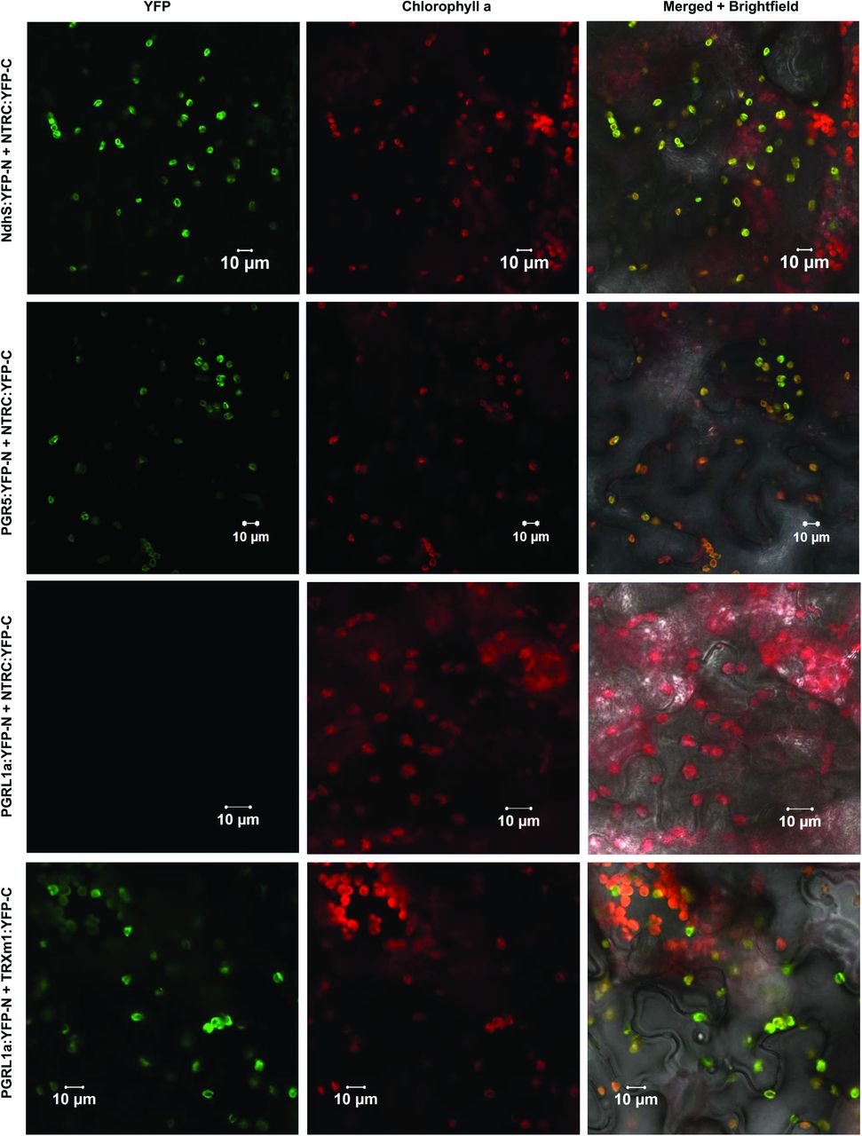
The potential interactions of NTRC with NdhS and PGR5 were further supported by positive results in bimolecular fluorescence complementation tests (BiFC). Co-expression of NTRC with both NdhS and PGR5 in Nicotiana benthamiana leaves resulted in YFP fluorescence that was strictly co-localized with chlorophyll autofluorescence, suggesting that it originated from the thylakoid membrane (Fig. 10). TRX-m1 interacted with PGRL1, while no interaction capability was detected between PGRL1 and NTRC (Fig. 10). Neither NdhS nor PGR5 interacted with TRX-x in BiFC, which was tested as a control (Suppl. Fig. S4).

Bimolecular fluorescence complementation (BiFC) tests of in planta interactions between chloroplast TRXs and potential target proteins in cyclic electron flow.
The left panel shows yellow fluorescent protein (YFP) fluorescence in green, the middle panel Chlorophyll a autofluorescence in red and the right panel a merged image of YFP, chlorophyll and brightfield images. YFP-N and YFP-C indicate expression of a fusion proteins including the N-terminal and C-terminal parts of YFP, respectively, in tobacco (Nicotiana benthamiana) leaves.
To assess the potential of the CEF-related proteins identified by Co-IP/MS (Table 1) and BiFC (Figure 10) to be targets of redox-regulation by TRXs, their amino acid sequences were analyzed for conserved cysteine residues. Of the NDH subunits identified as putative NTRC targets by Co-IP/MS, NdhS, NdhH, and Ndh48 contain cysteine residues that are conserved in angiosperms (Table 1, Suppl. Table S3, Suppl. Table S4, Suppl. Table S5), and therefore could in theory be subject to thiol regulation. NdhO and NdhU do not contain conserved cysteine residues, but they likely co-precipitate with the NTRC antibody because of their interactions with NdhH and NdhS subunits of the NDH complex, respectively (Shikanai, 2016).
PGR5 has been shown to form redox-dependent heterodimers with PGRL1 which have been proposed to be required for acceptance of electrons from ferredoxin and for reduction of PGRL1 (Hertle et al., 2013; Leister and Shikanai, 2013). The mature PGR5 polypeptide contains a single highly conserved cysteine residue (Munekage et al., 2002), which could hypothetically form an intermolecular disulfide with PGRL1 or some other partner, or be a target for S-nitrosylation or glutathionylation (Couturier et al., 2013; Zaffagnini et al., 2016). As direct determination of PGR5 redox state with the alkylation method was not feasible, we investigated if the redox state of PGRL1 is affected by NTRC deficiency or overexpression. PGRL1 contains six conserved thiol-sensitive cysteine residues that form inter- and intramolecular disulfides (Petroutsos et al., 2009; Hertle et al., 2013). We observed that PGRL1 was mostly oxidized in dark-adapted leaves, but underwent a transient reduction during approximately 60 seconds of illumination with growth light (Suppl. Fig. S3). This corresponds with the timescale of NPQ induction (Fig. 6), as well as with the transient increase in pmf and decrease in gH+ during dark-to-light transitions (Fig. 5). No significant difference to WT in PGRL1 reduction or protein content was detected in OE-NTRC (Suppl. Fig. S3, Fig. 3). PGR5 content of thylakoid membranes was, however, decreased in ntrc by 40% in comparison to WT (Fig. 3), in line with observations in a previous study (Yoshida and Hisabori, 2016).
DISCUSSION
The role of CEF around PSI in plant acclimation to fluctuating light conditions has attracted great attention during the past 10 years. Importance of CEF likely relies in its capacity to maintain redox balance in chloroplasts upon fluctuations in light intensity and during dark-to-light transitions (Yamori et al., 2016; Suorsa et al., 2016; Strand et al., 2016b). Plastidial thioredoxin systems, on the other hand, are crucial regulators of chloroplast metabolic reactions in the stroma. It has, however, remained unclear whether thioredoxin-related redox regulation is also involved in the attainment of CEF-mediated redox balance upon exposure of plants to changing light intensities. To this end, we applied here an in-vivo approach to investigate whether NTRC contributes directly to regulation of CEF pathways in chloroplasts.
Relevance of ntrc mutant in elucidating thioredoxin-dependent regulation of photosynthesis
The photosynthetic parameters measured for the ntrc knockout plants were not always in line with the results obtained with NTRC overexpressing plants (Fig. 2 and Fig. 4). In short photoperiod the ntrc plants have a highly pleiotropic phenotype (Fig. 3, Supl. Fig. 2) (Kirchsteiger et al., 2009; Pulido et al., 2010; Thormählen et al., 2015; Naranjo et al., 2016 Nikkanen et al., 2016; Pérez-Ruiz et al., 2017) that complicates the interpretation of results from the ntrc line. The high, slow-rising PIFR and increased dark-reduction of PQ in short-day grown ntrc (Fig. 2 and Fig. 3) were likely caused by a high NPQ (Fig. 8 and Suppl. Fig. S2) and its relaxation in darkness, lowered PSI content (Fig. 3) (Thormählen et al., 2015), and impaired stromal redox metabolism (Nikkanen et al., 2016; Pérez-Ruiz et al., 2017). Furthermore, we observed increased accumulation of reduced Fd, the substrate of the NDH complex, in ntrc (Fig. 2), which may be due to lower activity of CBC enzymes and consequent lower consumption of NADPH in carbon fixation (Nikkanen et al., 2016). Accordingly, a high NADPH/NADP+ ratio has been shown to activate CEF (Breyton et al., 2006; Okegawa et al., 2008). A high NADPH/NADP+ ratio together with high accumulation of reduced Fd may also explain the activation of NDH-dependent CEF by H2O2 (Strand et al., 2015; Strand et al., 2017), since oxidative treatment decreases the activation of redox-regulated CBC enzymes, and consequently the consumption of NADPH in illuminated chloroplasts. Accordingly, the ntrc mutant has a higher accumulation of H2O2 than WT (Suppl. Fig. S2 and Pulido et al., 2010), a higher NADPH/NADP+ ratio (Thormähälen et al., 2015), and lower activation states of CBC enzymes (Nikkanen et al., 2016; Pérez-Ruiz et al., 2017), which likely all contribute to the altered PIFR observed here. The PIFR was significantly diminished in ntrc plants grown in a longer photoperiod (Suppl. Fig. S2), where NPQ is lower (Suppl. Fig. S2) and the phenotype of ntrc has been shown to be less severe in terms of growth, chlorophyll content and efficiency of photochemistry (Lepistö et al., 2009; Lepistö et al., 2013). In ntrc electrons also accumulate in the PQ pool upon increases in light intensity (Fig. 6 and Fig. 8), although PSI is limited on the donor side (Fig. 6 and Fig. 9), indicating that electron transfer is limited between the PQ pool and PSI, possibly at Cyt b6f. Higher content of PTOX (Fig. 3) may assist to relax excitation pressure in the PQ pool of ntrc plants.
The pleiotropy described above for ntrc complicates the general interpretation of the results and makes it difficult to assess the contribution of NTRC to the regulation of photosynthesis when using the ntrc line. To avoid these obstacles we have constructed the OE-NTRC line, whose phenotype and development are not considerably dissimilar to WT (Toivola et al., 2013, Nikkanen et al., 2016) OENTRC in different backgrounds (ntrc, ndho and pgr5) provides a more reliable platform to examine the direct effects of NTRC on specific plastidial processes.
NTRC in regulation of CEF
In-depth analysis of NTRC overexpression lines in respect to thylakoid functional properties provided strong evidence that NTRC indeed is involved in the regulation of CEF in the thylakoid membrane. Compelling support for NTRC-induced activation of the NDH complex was obtained by analyzing thylakoid CEF-related functions in NTRC overexpression lines made on the backgrounds of ndho (OE NTRC ndho) and pgr5 (OE NTRC pgr5), incapable of performing NDH- and PGR-dependent CEF, respectively. Distinct effect of NTRC overexpression on the post-illumination rise of chlorophyll a fluorescence, the redox state of the plastoquinone pool in darkness as well as on the generation of the pmf and oxidation of P700 upon dark-to-light transitions and sudden increases in light intensity demonstrated the activating effect of NTRC on NDH-dependent CEF (Fig. 2, Fig. 3, Fig. 5, Fig. 6, and Fig 7). Higher activity of NDH-dependent CEF in plants overexpressing NTRC was shown not to be due to increased accumulation of the NDH complex or its substrate, because no increase in the accumulation of either NDH subunits or reduced Fd was detected in OE-NTRC plants in comparison to WT (Fig. 2 and Fig. 3). Furthermore, evidence for a direct effect of NTRC on the activation of NDH was obtained by identification of NDH subunits in a close proximity of the ferredoxin binding site as potential NTRC interactors (Table 1, Fig. 10). Although several NDH subunits were detected by Co-IP/MS, most likely only one or few of these subunits are genuine NTRC targets. The others likely co-precipitate with the NTRC antibody due to reciprocal interactions of the NDH subunits on the stromal side of the thylakoid membrane (Shikanai, 2016; Peltier et al., 2016). Existence of a thiol-regulated component in the ferredoxin binding site would provide a mechanism for dynamic control of the ferredoxin:plastoquinone oxidoreductase activity of the complex in response to fluctuations in light conditions. Redox-regulation of the NDH complex would allow rapid adjustment of pmf and nonphotochemical quenching as well as a maintenance of redox balance between the electron transfer chain and the electron sink capacity of stromal acceptors, most importantly the CBC. In high light, less active NDH could prevent the reverse function of the complex (i.e. oxidization of PQ to reduce ferredoxin and transfer of H+ from lumen to stroma) in conditions of high pmf and a reduced PQ pool (Strand et al., 2017a).
Notably, the overexpression of NTRC also affects the function of PGR5. Nevertheless, as discussed below, the effect is not necessarily related to the putative role of PGR5 in CEF. Our results, more likely, support the hypothesis (Avenson et al., 2005; Tikkanen et al., 2015; Kanazawa et al., 2017; Armbruster et al., 2017) that PGR5 is involved in control of the proton conductivity of the thylakoid membrane that, consequently, affects the generation of pmf.
Regulation of pmf and the thylakoid redox balance via NDH and NTRC during changes in light conditions
Overexpression of NTRC caused elevated pmf under all light conditions, while no significant changes were observed in PSII quantum yield or thylakoid proton conductivity in comparison to WT (Fig. 5, Fig. 6, Fig. 7 and Fig. 8). These results strongly suggest that the elevation of pmf derives from enhanced CEF. Increased P700 oxidation during dark-to-light transitions in OE-NTRC was fully reverted in OE-NTRC ndho, and a lack of NDH also delayed the ability to oxidize P700 during the high light phases in fluctuating light (Fig. 6, Fig. 9). It is therefore evident that the NDH complex regulates the trans-thylakoid pmf as well as the redox balance between the electron transfer chain and the stroma, and this regulation is under the control of the stromal TRX systems, with our results suggesting a specific role for the NTRC system.
While our results demonstrate enhancement of the NDH-dependent CEF by NTRC overexpression (Fig. 2), earlier studies have revealed an inhibition of NDH-dependent CEF upon TRX-m4 overexpression and, conversely, an enhancement in trxm4 mutants (Courteille et al., 2013). Thus, it is conceivable that the two chloroplast TRX systems regulate CEF in an antagonistic way, although it remains to be elucidated how such regulation might be mechanistically accomplished. We propose that in low light and upon sudden changes in the light intensity, NTRC is crucial for activation of the NDH-dependent CEF, while the TRX-m4-dependent inhibition of NDH-CEF requires higher light intensity or longer duration of illumination. Moderate to high-light illumination is required to fully activate the FTR-dependent TRX system (reviewed in Geigenberger et al. 2017) that possibly contributes to downregulation of NDH. In OE-NTRC the NDH-dependent CEF is constitutively active in light, which contributes to elevated pmf in all light intensities. Upon transition from dark to low light, there is less difference between OE-NTRC and WT in terms of pmf formation (Fig. 7), because in those conditions the NTRC-mediated activation of NDH occurs similarly in WT and OE-NTRC.
The NDH complex translocates protons from the stroma to the lumen not only via Cyt b6f, but also itself functions as a proton pump with a 2 H+/ e− stoichiometry (Strand et al., 2017a). NDH-mediated CEF therefore contributes relatively more to pmf generation and consequently to ATP synthesis and NPQ induction than the PGR-dependent pathway. It has been postulated that the NDH complex is unlikely responsible for CEF during the early induction phase of photosynthesis, due to a low concentration of the complex in thylakoids in relation to the total PSI content (Joliot and Joliot, 2002). However, the NDH complex forms functional CEF-supercomplexes with PSI in stroma thylakoids (Peng et al., 2008), and a single NDH complex can bind up to six PSI complexes (Yadav et al., 2017), indicating that even a relatively low NDH content may have a significant impact on pmf generation.
Contribution of TRX systems to PGR-related CEF pathway
Increased activation of NDH-CEF is not alone sufficient to explain all observed changes of pmf in OENTRC plants. When compared to WT, pmf remained elevated in OE-NTRC ndho during the first seconds of photosynthetic induction and at steady state in growth and high light (Fig. 5 and Fig. 7). These results could be explained by activation of PGR-dependent CEF as well in plants overexpressing NTRC. Stromal thiol redox state has been previously suggested to control the PGR-dependent CEF by a component that has a midpoint redox potential of −310 mV (Hertle et al., 2013; Strand et al., 2016a). It has also been proposed that m-type TRXs, with redox potentials between −357 and −312 mV (Collin et al., 2003; Yoshida et al., 2015), reduce an intermolecular disulfide in PGRL1 homodimers, and subsequently, the released monomeric PGRL1 may function as the ferredoxin-plastoquinone reductase (Hertle et al., 2013). Here we confirm the previously reported transient reduction of PGRL1 during dark-to-light transitions (Hertle et al., 2013), but NTRC overexpression does not intervene in the reduction (Suppl. Fig. S3). Moreover, TRX-m1 but not NTRC interacts with PGRL1 in BiFC (Fig. 10). Our results thus support the hypothesis that TRX-m is a primary reductant of PGRL1. Crosstalk between NTRC and FTR-dependent systems (Toivola et al., 2013; Thormählen et al., 2015; Nikkanen et al., 2016), and the interaction of NTRC with TRX-m1 in BiFC (Nikkanen et al., 2016), further support the interpretation that the activation of PGR-dependent CEF is indirectly increased in NTRC-overexpressing plants through enhancement of TRX-m reduction. This would also be in line with the steady state pmf increase observed in OE-NTRC ndho in comparison to WT (Fig. 5 and Fig. 7).
Alternatively, NTRC overexpression may affect the function of PGR5 in a way that is independent of its involvement in CEF. Redox regulation of PGR5 may occur to control its association with the ATP synthase during dark-to-light and low-to-high light transitions, and thereby allows inhibition of the ATP synthase in an unknown mechanism, as suggested earlier (Avenson et al., 2005; Tikkanen et al., 2015; Kanazawa et al., 2017; Armbruster et al., 2017). Such a mechanism would result in acidification of the lumen and induction of NPQ, allowing dissipation of excess excitation energy from the electron transfer chain until CBC is activated. This hypothesis is supported by the impaired abilities of pgr5 and OE-NTRC pgr5 to control the activity of the ATP synthase at early stages of dark-light transitions and upon transitions to high light intensities (Avenson et al., 2005, Fig. 5 and Fig. 7). Furthermore, the elevated NTRC content in leaves caused decreased thylakoid proton conductivity upon increases in light intensity (Fig. 7), suggesting that NTRC controls the PGR5-dependent down-regulation of proton efflux from lumen. This is supported by the identification of PGR5 as a potential NTRC interactor (Table 1, Fig. 3, Fig. 10).
The initial strong pmf increase in OE-NTRC after onset of growth light illumination was evident also in both the OE-NTRC ndho and OE-NTRC pgr5 plants (Fig. 5) indicating that this pmf peak is not caused by CEF. More likely, the initial pmf results from dark-activation of the CBC enzymes in plants overexpressing NTRC (Nikkanen et al. 2016), which provides an enhanced ability of the stroma to accept electrons from the PETC upon dark-to light transition and consequently enhances proton pumping to the lumen.
Cooperative regulation of photosynthetic electron transfer and carbon fixation by chloroplast thioredoxin systems
Light-dependent reductive activation of the ATP synthase, the CBC and the NADP-malate dehydrogenase (NADP-MDH) by TRXs has been well established for several decades (reviewed in (Buchanan, 2016). More recently, knowledge of TRX-mediated control has been extended to various regulatory and photoprotective mechanisms of photosynthesis, including regulation of state transitions (Rintamäki et al., 2000; Shapiguzov et al., 2016), NPQ (Hall et al., 2010; Brooks et al., 2013; Naranjo et al., 2016; Da et al., 2017) and CEF (Courteille et al., 2013; Hertle et al., 2013; Strand et al., 2016a). We propose here a model, comprising a cooperative function of the two chloroplast TRX systems with distinct reductants and redox potentials that allows the maintenance of redox balance between the two photosystems and stromal metabolism during fluctuations in light conditions. This is achieved through dynamic regulation of the activities of the ATP synthase, NPQ, the NDH complex, PGRL1/PGR5 as well as the LHCII kinase STN7 by reversible thiol modifications. We propose a specific role for NTRC in regulating NDH-CEF, the ATP synthase and CBC enzymes in low light, dark-to-light transitions and during sudden increases in light intensity, as schematically depicted in Fig. 11.

A schematic model of the role of chloroplast TRX systems in regulating CEF and the pmf during dark-to-light transitions and fluctuations in light intensity.
(A–C) Dark-adapted leaves (A), transition from dark to low light (B) and transition from low to high light (C). Blue color indicates the approximate reduction levels of different photosynthetic redox components based on data in the current paper (Figures 1, 2, 8, and 9) as well as other reports (Yoshida and Hisabori, 2015; Nikkanen et al., 2016; Schreiber, 2017; Schreiber et al., 2017). Green and red arrows indicate activating and inhibitory effects, respectively, while orange color represents the thiol regulation by NTRC and purple by the Fd-TRX system. Thicker lines depict stronger effect than thin and dotted lines. For details see the text. The arrow to NPQ refers to induction of the qE component of NPQ due to acidification of the lumen (Demmig-Adams et al., 2012). The arrows representing reduction of PQ by the NDH complex and PGRL1 have been drawn through the lumen only to increase clarity of the illustration.
In darkness, a proportion of NDH complexes in the thylakoid membrane is activated by NTRC, and moderate chlororespiration from NDH to PTOX occurs. Due to an inactive ATP synthase and proton pumping activity of NDH, a weak proton gradient over the thylakoid membrane is established. Redox-regulated CBC enzymes are inactive, causing PC and P700 to be reduced (Schreiber, 2017) due to lack of electron acceptors in the stroma. In OE-NTRC, chlororespiration via NDH to the PQ pool is elevated due to increased amount of active NDH complexes. This leads to increased protonation of the lumen and higher reduction of the PQ pool. Proportions of the ATP synthase and CBC enzyme pools are activated due to high NTRC content.
Upon transition from dark to low light, the ATP synthase pool is fully and the CBC enzyme pool partially reduced by NTRC in WT plants. Delay in activation of the CBC enzymes causes, however, reduction of the PQ pool due to scarcity of stromal acceptors that limits electron transfer. NTRC contributes to activation of NDH-dependent CEF, which alleviates electron pressure at PSI and transiently increases pmf and induces NPQ. In OE-NTRC, P700 and PC are effectively oxidized upon onset of low illumination, as the acceptor side limitation is negligible due to fully active NDH-dependent CEF, ATP synthase and redox-activated CBC enzymes. This results in an elevated ΔpH and faster induction of NPQ in comparison to WT at the initial phase of illumination. At steady state NPQ is lower than in WT despite of high ΔpH, suggesting downregulation of NPQ by thioredoxin via a ΔpH-independent mechanism, as reported previously by Brooks et al. (2013).
When a leaf is shifted from low to high irradiance, both TRX systems become fully active, and the CBC enzymes as well as the PGR-dependent CEF are fully activated. NTRC affects PGR5-dependent inhibition of the ATP synthase, which contributes to accumulation of protons in the lumen. Consequently, NPQ and down-regulation of electron transfer at Cyt b6f are induced. Electrons are effectively pulled from PSI, and the donor side is limiting electron transfer. In OE-NTRC, increased reduction of PGR5 likely leads to stronger down-regulation of the ATP synthase. This, together with proton pumping by constantly active NDH and possibly through increased TRX-m-mediated activation of the PGR-dependent pathway, results in high pmf. NPQ is however lower than in WT due to ΔpH-independent downregulation of NPQ by overexpressed NTRC.
CONCLUSIONS
In the present paper, we have shown that the chloroplast NADPH-dependent thioredoxin system (NTRC) stimulates cyclic electron flow around PSI by activating the thylakoid NDH complex. We propose that NTRC-mediated activation of CEF is particularly important during fluctuations in light intensity and in low light in order to keep redox balance between photosynthetic electron transfer chain in the thylakoid membranes and stromal metabolism. Interactions assays suggest that a TRX-target exists close to the ferredoxin binding and oxidation site of the NDH-complex, and possibly involves the NdhS subunit. We further suggest that NTRC regulates photosynthetic redox poise by promoting PGR5-dependent downregulation of the activity of the ATP synthase upon transitions from low to high light intensity. This results in acidification of the lumen, which is needed for induction of photoprotective mechanisms.
MATERIALS AND METHODS
Plant material and growth conditions
Experiments have been done with Arabidopsis thaliana wild type (WT) lines of the Columbia ecotype (Col-0 and Col-gl1), and with the following transgenic lines: NTRC overexpression line (Toivola et al., 2013), T-DNA knockout mutants of NTRC (At2g41680, SALK_096776) (Lepistö et al., 2009), ndho (At1g74880, SALK_ 068922) (Rumeau et al., 2005) and STN7 (AT1G68830, SALK_073254) (Bellafiore et al., 2005) as well as the pgr5 mutant (AT2G05620) (Munekage et al., 2002). The plants were grown in a photoperiod of 8 h light / 16 h darkness at 23 °C under 200 μmol of photons m−2◻s−1 for all experiments except for the measurements shown in Suppl. Fig. S2, for which plants were grown in a 12 h /12 h photoperiod under 130 μmol m−2◻s−1. Wild type tobacco (Nicotiana benthamiana) plants used in BiFC tests were grown under 130◻μmol photons m−2◻s−1 at 23◻°C in a 16◻h light/8◻h dark photoperiod. The OE-NTRC ndho and OE-NTRC pgr5 lines were generated by Agrobacterium tumefaciens and floral dipping –mediated transformation of the ndho knockout and pgr5 mutant lines, respectively, with the NTRC overexpression construct as described previously (Toivola et al., 2013). The OE-NTRC ndho and OE-NTRC pgr5 plants used in the experiments were heterozygous T2 generation plants that were selected on agar plates with 0.5X Murashige-Skoog medium (MS) (Murashige and Skoog, 1962) and 50 μg/ml kanamycin. The plants were subsequently transferred to soil and grown in a 8 h light / 16 h darkness photoperiod at 23 °C under 200 μmol of photons m−2◻s−1 for four weeks before usage in the experiments. As control, OE-NTRC plants were similarly selected on kanamycin-containing plates while WT Col-0 and WT Col-gl1 (ecotype of the pgr5 mutant) plants were grown on 0.5X MS-agar plates without antibiotics for an equivalent time.
Determination of H2O2 content in leaves
The hydrogen peroxide content in leaves was estimated by staining with diaminobenzidine (DAB), as previously described in Lepistö et al. (2013). Detached leaves from 4-weeks-old WT, ntrc and OENTRC plants were incubated overnight in darkness in 0.1 mg ml−1 solution of diaminobenzidine (DAB; Sigma-Aldrich) (pH 3.8), after which the leaves were illuminated with either 40 or 200 μmol photons m−2◻s−1 for 1h. Chlorophyll was then bleached by incubating the leaves in ethanol and subsequently photographed. Image J software (Schneider et al., 2012) was used to quantify the intensity of the staining.
Measurement of Chlorophyll a fluorescence and P700 and Fd redox changes
The post-illumination chlorophyll a fluorescence rise (PIFR) was measured from detached leaves with the Multicolor-PAM fluorometer (Walz). A 480 nm measuring beam at an intensity of 0.2 μmol photons m−2◻s−1 was used to measure fluorescence changes after illumination of dark-adapted (30 min) leaves with 67 μmol photons m−2◻s−1 of white actinic light for 500 seconds, with saturating pulses of 800 ms (10000 μmol photons m−2◻s−1) in the beginning and at 400 s to determine Fm and Fm’. The actinic light was then switched off and the changes in chlorophyll a fluorescence in the dark were observed for 300 s. A 10 s pulse of far red light was then given to fully oxidize the PQ pool, and the subsequent re-reduction PQ pool was detected through a rise in Chl fluorescence.
The OJIP transients were recorded with the Multicolor-PAM from dark-adapted (30 min) leaves and from leaves pre-illuminated with far red light (intensity setting 15) for 6 s, according to the method described by (Toth et al., 2007). A saturating pulse of 3000 μmol photons m−2◻s−1 and measuring light at 440 nm were used in the measurements.
The Dual-PAM-100 was used to simultaneously record the Chl a fluorescence and P700-dependent difference in absorbance at 875 and 830 nm during transitions from dark to 166 μmol photons m−2 s−1 (Fig. 6) and during a light regime, where a 620 nm AL fluctuates between 39 and 825 μmol photons m−2 s−1 (Fig. 8 and Fig. 9). Saturating pulses were administered at 10 or 15 s intervals for the measurements in Fig. 6 and at 15 s intervals for the first minute after onset of illumination, and at 20 s intervals thereafter for Fig. 8 and Fig. 9. Because ntrc leaves are very small in size and low in chlorophyll content, it was in some cases necessary to record from two or three leaves simultaneously to obtain a P700 signal of sufficient quality. The parameters shown were calculated with the Dual-PAM-100 software as described by Bilger and Björkman (1990), Klughammer and Schreiber (2008a, 2008b) and Kramer et al., (2004).
For determination of Fd redox state, the Dual/Klas-NIR (Walz) spectrometer was used to record the four absorbance differences between 785 and 840, 810 and 870, 870 and 970, as well as 795 and 970 nm, from which the redox changes of P700, PC and Fd were deconvoluted as described by Klughammer and Schreiber (2016) and Schreiber (2017). A similar illumination and post-illumination regime was used as described above for the measurement of PIFR, with the exception that dark-adapted leaves were illuminated with 61 μmol photons m−2 s−1 of 630 nm instead of white actinic light. Measured Fd redox changes were then normalized to the level maximal Fd reduction, which was determined according to Schreiber and Klughammer (2016).
Measurement of electrochromic shift (ECS)
In order to measure the magnitude and kinetics of pmf formation, changes in the electrochromic shift (ECS, P515) signal were recorded with the Dual-PAM-100 and the P515/535 accessory module (Walz) (Schreiber and Klughammer, 2008, Klughammer et al., 2013). A dual beam difference signal between 550 and 515 nm was used to avoid distortion of results by scattering effects. A measuring light at a 2000 Hz pulse frequency was used in all ECS measurements. For the dark-to-light and low-to-high light transition measurements in Fig. 5 and Fig. 7, plants were first dark-adapted for 30 min. A single-turnover saturating flash (20 μs) of 14000 μmol photons m−2s−1 was then applied to obtain ECSST, a maximum absorbance change value that was used to normalize all results to account for differences in leaf thickness and chlorophyll content between individual leaves and lines (Kramer and Crofts, 1989). The obtained values of ECSST were in good correlation with the differences in chlorophyll content in OE-NTRC and ntrc lines reported previously (Toivola et al., 2013). In order to distinguish the light-induced ECS change (ECST) from signal drift and baseline change, dark intervals of 250 ms were applied at the following time points after the onset of AL illumination: 0.8; 2.7; 4.7; 6.7; 8.7; 10.7; 12.7; 16.7; 20.7; 24.7; 28.7; 32.7; 36.7; 40.6; 44.7; 48.7; 52.7; 56.7; 60.7; 80.6; 100.5; 120.5; 140.5 and 160.5 s after onset of illumination. Additionally, during the shift from low to high irradiance (Fig. 7), dark intervals were applied at 1.1; 5.1; 9.1; 13.1; 23.1; 33.1; 43.1; 53.1; 73.1; 93.1; 113.1; 133.1 and 153.1 s after the increase in light intensity. ECST was calculated as the difference between total ECS in light and an Y0 value obtained from the first-order exponential fit to the decay kinetics of the ECS signal during a dark interval. Total pmf was then calculated as ECST/ECSST. The gH+ parameter, describing thylakoid membrane conductivity to protons, was calculated as the inverse of the time constant of a first-order exponential fit to ECS decay kinetics during a dark interval (Cruz et al., 2001; Avenson et al., 2005; Cruz et al., 2005). Partitioning of total pmf to its components ΔpH and ΔΨ was determined from the light-off response of the ECS signal (Cruz et al., 2001) after 3 min illumination, also using the Dual PAM ECS module as described by Schreiber and Klughammer (2008). Same settings were used for determination of pmf partitioning as for the dark-to-light and low-to-high light transition measurements.
Protein extraction, alkylation of thiols and SDS-PAGE
Proteins and thylakoids were isolated as previously described (Lepistö et al., 2009), while chlorophyll content was determined according to (Porra et al., 1989) and protein content with the Bio-Rad Protein Assay kit. For determination of the redox states of TRX-regulated proteins, leaf proteins were precipitated with trichloroacetic acid (TCA) and free thiols in proteins alkylated with N-ethylmaleimide (NEM, Sigma-Aldrich). After alkylation protein disulfides were reduced with dithiothreitol (DTT, Sigma-Aldrich) and subsequently produced thiols were alkylated with methoxypolyethylene glycol maleimide Mn 5000 (MAL-PEG, Sigma-Aldrich) as described earlier (Nikkanen et al., 2016). Sodium dodecyl sulfate polyacrylamide gel electrophoresis (SDS-PAGE) and immunoblotting was performed as reported in (Nikkanen et al., 2016). For running the MAL-PEG samples pre-cast 4-20% Mini-PROTEAN TGX gels (Bio-Rad) were used, except for the gel in Fig. 1B, Suppl. Fig. S1A and Suppl. Fig. S3A, where a 12% polyacrylamide gel was used. PVDF membranes were probed with antibodies raised against NTRC (Lepistö et al., 2009), D1 (Research Genetics, Inc (Thermo Fisher)), PsaB (Agrisera, AS10 695), Cyt f (kindly provided by L. Zhang), PTOX (kindly provided by M. Kuntz), NdhH (Agrisera), NdhS (Agrisera), CF1γ (Agrisera, AS08 312), PGRL1 (Agrisera, AS10 725), PGR5 (Agrisera) or phosphothreonine (P-Thr) (New England Biolabs). Membranes were then treated with a horseradish peroxidase (HRP)-conjugated goat anti-rabbit secondary antibody (Agrisera, AS09 602) for 2◻h. All immunoblots shown are representative of at least three biological replicates. Quantifications of protein content shown in Fig. 3 were performed using the ImageJ software (Schneider et al., 2012) and normalized according to the intensity of Li-Cor Revert Total Protein Stain. Statistical significance was determined using two-tailed Student’s T-tests for unequal variances with p-values below 0.05 interpreted as statistically significant.
Co-immunoprecipitation and Mass spectrometry
For co-immunoprecipitation (Co-IP), WT, ntrc and OE-NTRC leaves were frozen in liquid N2, lysed in Pierce IP Lysis buffer containing 1 % NP-40 detergent (Thermo-Fisher), and immunoprecipitated in a resin containing NTRC-specific antibody using the Pierce Co-IP kit (Thermo-Fisher) with an affinity-purified NTRC-specific antibody, as described previously (Nikkanen et al., 2016). Co-IP eluates were denatured and purified by SDS-PAGE in a 6% acrylamide gel with 6 M urea, subjected to in-gel tryptic digestion and the extracted peptides analyzed with the Q Exactive Hybrid Quadruple-Orbitrap mass spectrometer (Thermo-Fisher Scientific) in DDA mode as previously described (Trotta et al., 2016). MS/MS spectra were analyzed with an in-house installation of Mascot (v.2.4) (Matrix Science) search engine and analyzed with Proteome Discoverer (v.1.4) Software (Thermo Scientific), restricting the search to the non-redundant database TAIR10 supplemented with most common laboratory contaminants (Trotta et al., 2016). Peptides were validated by Decoy Database Search, with target false discovery rates (FDR) set to be below 0.01 (strict) or below 0.05 (relaxed).
BiFC tests
Bimolecular fluorescence complementation tests (BiFC) were performed as described in (Nikkanen et al., 2016). For the current study, coding sequences of PGR5, PGRL1a and NdhS obtained from Arabidopsis Biological Resource Center (ABRC) were cloned into pSPYNE-35S and pSPYCE-35S binary vectors (Walter et al., 2004), and the resulting constructs were checked by sequencing. Primer sequences used for cloning are listed in Suppl. Table S6. Imaging of YFP and chlorophyll autofluorescence from N. benthamiana leaves infiltrated with Agrobacterium tumefaciens strain GV3101 carrying the appropriate binary vectors was performed with a Zeiss LSM780 laser scanning confocal microscope at 3 days after infiltration. The negative result between PGRL1:YFP-N and NTRC:YFP-C also serves as a negative control.
Multiple alignment of amino acid sequences
Amino acid sequences of NdhH, Ndh48, NdhS, NdhJ and Ndh45 in Arabidopsis thaliana and, as available, in Populus trichocarpa, Vitis vinifera, Glycine max, Solanum lycopersicum, Oryza sativa, Sorghum bicolor, Brachypodium distachion, Physcomitrella patens, Selaginella moellendorffii and Synechocystis PCC 6803 were obtained from the UniProtKB database and aligned with the Clustal Omega 1.2.4 online alignment tool (Sievers et al., 2011) using default settings.
Accession Numbers
The Arabidopsis Genome Initiative locus identifiers (AGI) used in this paper are listed in Table 1, Suppl. Table S2, Suppl. Table S3, Suppl. Table S4, Suppl. Table S5, Suppl. Table S6 and Suppl. Dataset 1.
Author contributions
L.N. and E.R. designed the research, L.N., J.T., and A.T. performed the research, L.N., J.T., A.T., M.T., and E.R. analyzed the data, L.N. and E.R. wrote the article with input from E-M.A., M.T., A.T., M.G.D., and J.T.
Funding information
This work was funded by the Academy of Finland Grants 276392 (to E.R.) and 307335 (the Center of Excellence in Molecular Biology of Primary Producers to E-M.A.) and by the Doctoral Program in Molecular Life Sciences in the University of Turku Graduate School (to L.N.).
ACKNOWLEDGEMENTS
We thank Jesse Ojala for assistance with the experiments, Esa Tyystjärvi for the advice for PIFR measurements, Sari Järvi and Marjaana Suorsa for advice on the use of antibodies, Alexandrina Stirbet for expert advice on measurement of OJIP transients, and Mika Keränen, Kurt Ståhle and Tapio Ronkainen for technical assistance.
LITERATURE CITED


